Japanese
English
- 有料閲覧
- Abstract 文献概要
- 1ページ目 Look Inside
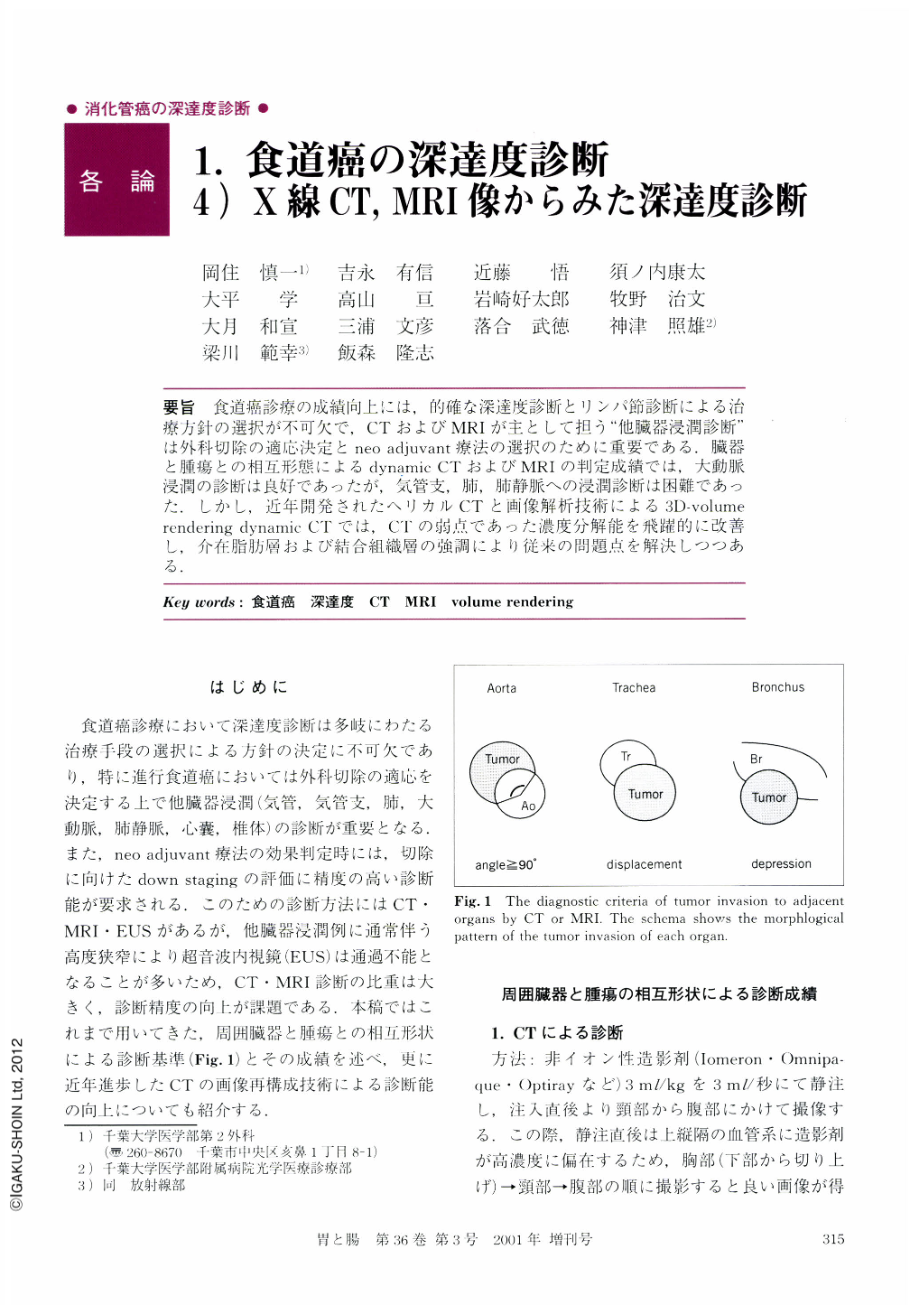
要旨 食道癌診療の成績向上には,的確な深達度診断とリンパ節診断による治療方針の選択が不可欠で,CTおよびMRIが主として担う“他臓器浸潤診断”は外科切除の適応決定とneo adjuvant療法の選択のために重要である.臓器と腫瘍との相互形態によるdynamic CTおよびMRIの判定成績では,大動脈浸潤の診断は良好であったが,気管支,肺,肺静脈への浸潤診断は困難であった.しかし,近年開発されたヘリカルCTと画像解析技術による3D-volume rendering dynamic CTでは,CTの弱点であった濃度分解能を飛躍的に改善し,介在脂肪層および結合組織層の強調により従来の問題点を解決しつつある.
Diagnosis of the depth of a tumor in case of esophageal cancer is very important for the determination of the therapeutic course. The diagnosis of the invasion to the adjacent organ is especially necessary for deciding whether surgical resection of the tumor or neo adjuvant therapy is indicated. The conventional methods of diagnosis using CT or MRI depend on the morphological relation between the tumor and the adjacent organ, but these methods are almost ineffective for diagnosing invasion of the bronchus, lung, or pulmonary vein. The recent advance of high speed and high resolution helical CT and the volume rendering technique enables us to visualize the fat layer or fibrous layer between the tumor and the adjacent organ. Because of this the diagnosis of the invasion of a tumor to the bronchus, lung, or pulmonary vein has become much easier.

Copyright © 2001, Igaku-Shoin Ltd. All rights reserved.


