Japanese
English
- 有料閲覧
- Abstract 文献概要
- 1ページ目 Look Inside
- サイト内被引用 Cited by
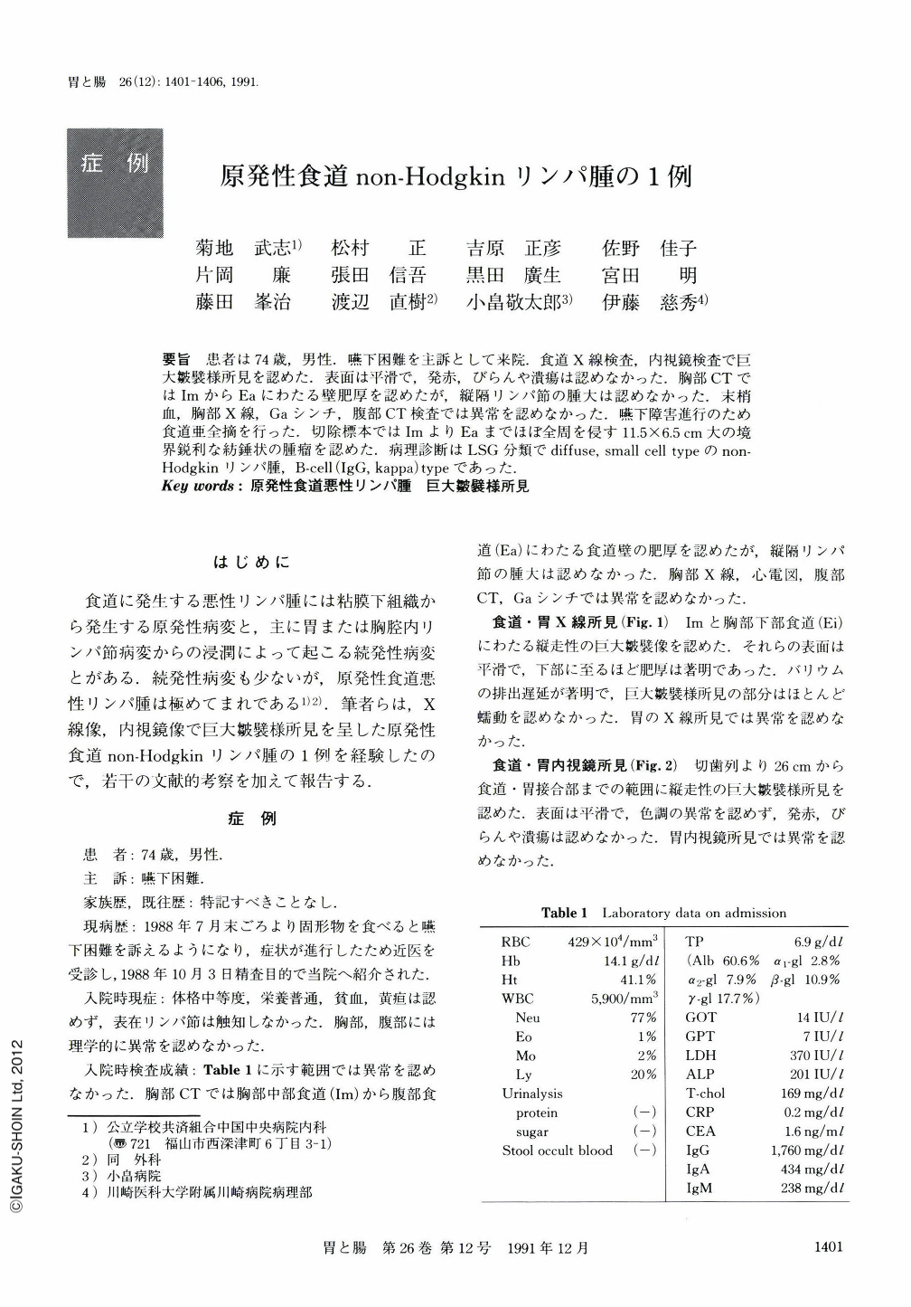
要旨 患者は74歳,男性.嚥下困難を主訴として来院.食道X線検査,内視鏡検査で巨大皺襞様所見を認めた.表面は平滑で,発赤,びらんや潰瘍は認めなかった.胸部CTではImからEaにわたる壁肥厚を認めたが,縦隔リンパ節の腫大は認めなかった.末梢血,胸部X線,Gaシンチ,腹部CT検査では異常を認めなかった.嚥下障害進行のため食道亜全摘を行った,切除標本ではImよりEaまでほぼ全周を侵す11.5×6.5cm大の境界鋭利な紡錘状の腫瘤を認めた.病理診断はLSG分類でdiffuse,small cell typeのnon-Hodgkinリンパ腫,B-cell(IgG,kappa)typeであった.
Reported was a 74-year-old male patient visiting our hospital with the chief complaint of dysphagia. Giant folds were observed in a wide area from the middle intra-thoracic esophagus (Im) to the lower intrathoracic esophagus (Ei) as revealed by x-ray examination (Fig. 1). By endoscopy, the above finding was confirmed in the area 26 cm distal to the incisors downward. The mucosal surface of the lesion was smooth, with no redness, erosion or ulce (Fig. 2). Reactive lymphoreticular hyperplasia was diagnosed histologically by biopsy. Preoperative examination revealed no abnormality in peripheral blood, thoracic x-ray, Ga scintigram or abdominal CT scanning. Because of increasing dysphagia, subtotal resection of the esophagus was performed. The resected specimen showed a well-defined fusiform tumor, measuring 11.5×6.5 cm in size, from the middle intra-thoracic esophagus (Im) down to the abdominal esophagus (Ea), involving almost entire circumference (Fig. 3). The final pathological diagnosis was diffuse small-cell type non-Hodgkin's lymphoma comprising B-cells (IgG, kappa) as classified by LSG classification(Figs. 4~6).

Copyright © 1991, Igaku-Shoin Ltd. All rights reserved.


