Japanese
English
- 有料閲覧
- Abstract 文献概要
- 1ページ目 Look Inside
- 参考文献 Reference
- サイト内被引用 Cited by
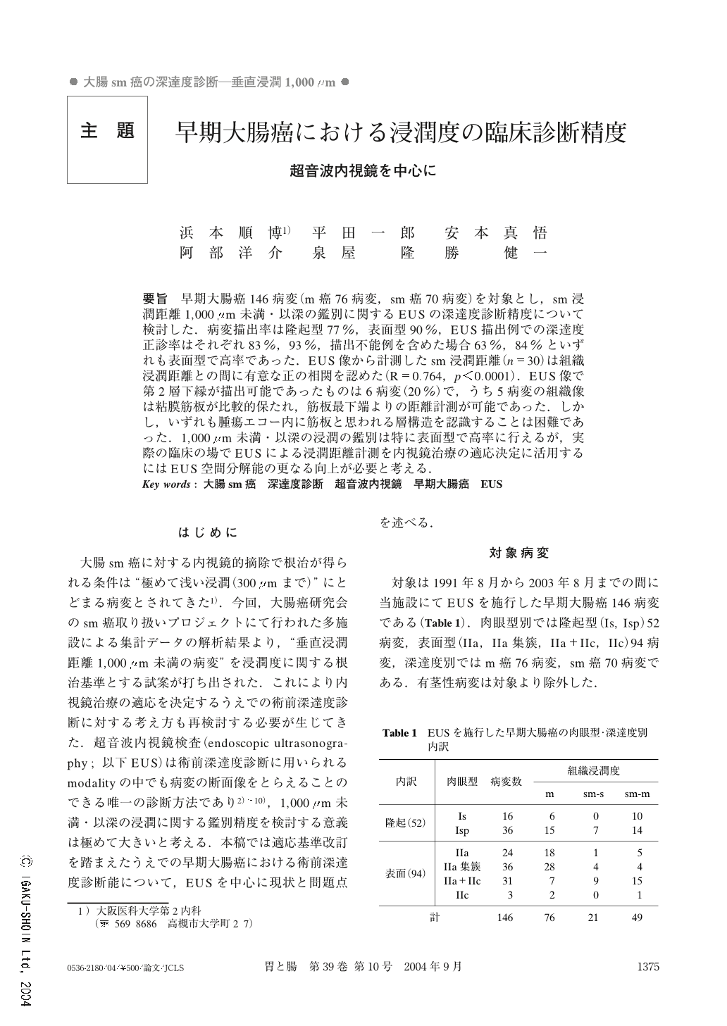
要旨 早期大腸癌146病変(m癌76病変,sm癌70病変)を対象とし,sm浸潤距離1,000μm未満・以深の鑑別に関するEUSの深達度診断精度について検討した.病変描出率は隆起型77%,表面型90%,EUS描出例での深達度正診率はそれぞれ83%,93%,描出不能例を含めた場合63%,84%といずれも表面型で高率であった.EUS像から計測したsm浸潤距離(n=30)は組織浸潤距離との間に有意な正の相関を認めた(R=0.764,p<0.0001).EUS像で第2層下縁が描出可能であったものは6病変(20%)で,うち5病変の組織像は粘膜筋板が比較的保たれ,筋板最下端よりの距離計測が可能であった.しかし,いずれも腫瘍エコー内に筋板と思われる層構造を認識することは困難であった.1,000μm未満・以深の浸潤の鑑別は特に表面型で高率に行えるが,実際の臨床の場でEUSによる浸潤距離計測を内視鏡治療の適応決定に活用するにはEUS空間分解能の更なる向上が必要と考える.
We investigated the accuracy of endoscopic ultrasonography (EUS) in diagnosing the depth of invasion of carcinomas, especially when differentiating between invasive lesions less than 1,000μm deep and other deeper lesions. The subject consisted of 76 mucosal cancers and 70 submucosal invasive cancers.
The detection rate by EUS was 77% for the protruded type and 90% for the flat type. Regarding the lesions that could be detected by EUS, the diagnostic accuracy rate was 83% for the protruded type and 93% for the flat type, indicating that both rates were higher for the flat type.
We attempted to calculate the depth of submucosal invasion in EUS images (of 30 lesions). There was a significantly positive correlation between the depth of submucosal invasion as calculated in the EUS images and that verified in histologic specimen (R=0.764, p<0.0001). In 6 (20%) of these 30, the bottom of the second layer could be recognized in the tumor echo. In 5 of those 6 lesions, the muscularis mucosae was almost perfectly preserved and the depth of submucosal invasion was histologically calculated from the bottom of the muscularis mucosae. However, it was often difficult to confirm the layer corresponding to the muscularis mucosae in the EUS image.
EUS is useful in the differentiation between invasive lesions less than 1,000μm deep and other deeper lesions, especially in the flat type of early colorectal carcinoma. However, it is necessary to improve the resolution of EUS in order to calculate depth of invasion in the EUS image to decide whether endoscopic resectionis indicated or not.
1) Second Department of Internal Medicine, Osaka Medical College, Takatsuki, Japan

Copyright © 2004, Igaku-Shoin Ltd. All rights reserved.


