Japanese
English
- 有料閲覧
- Abstract 文献概要
- 1ページ目 Look Inside
- 参考文献 Reference
- サイト内被引用 Cited by
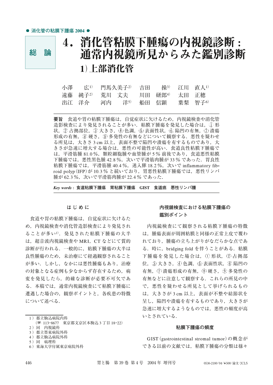
要旨 食道や胃の粘膜下腫瘍は,自覚症状に欠けるため,内視鏡検査や消化管造影検査により発見されることが多い.粘膜下腫瘍を発見した場合は,①形状,②占拠部位,③大きさ,④色調,⑤表面性状,⑥陥凹の有無,⑦潰瘍形成の有無,⑧硬さ,⑨多発性の有無などについて観察する.悪性を疑わせる所見は,大きさ3cm以上,表面不整で陥凹や潰瘍を有するものであり,大きさが急速に増大する場合は,悪性の可能性が高い.食道良性粘膜下腫瘍では,平滑筋腫81.0%,顆粒細胞腫や血管腫が5%前後であり,食道悪性粘膜下腫瘍では,悪性黒色腫42.8%,次いで平滑筋肉腫が33%であった.胃良性粘膜下腫瘍では,平滑筋腫40.4%,迷入膵18.2%,次いでinflammatory fibroid polyp(IFP)が10.3%と続いており,胃悪性粘膜下腫瘍では,悪性リンパ腫が62.3%,次いで平滑筋肉腫が22.4%であった.
As there are usually no symptoms associated with esophageal or gastric submucosal tumors, they are often found first by endoscopy or double-contrast radiographic examination. When a submucosal tumor is detected, it should be defined by its shape, region, size, color, solidity, surface appearance, presence of depressions, ulcers, or multiple lesions, etc. The findings of a size in excess of3cm, irregular surface, accompanied by depressions or ulcerations, and features of rapid growth represent frequently malignant manifestations of these lesions.
The cases examined in the present study were reported as leiomyoma (81.0%), granular cell tumor, and hemangioma (approximately 5%) as benign esophageal submucosal tumors, and malignant melanoma (42.8%) or leiomyosarcoma (33%) as malignant esophageal submucosal tumors. The tumors were identified as leiomyosarcoma (40.4%), aberrant pancreas (18.2%), or inflammatory fibroid polyp (IFP) (10.3%) as benign gastric submucosal tumors, and malignant lymphoma (62.3%) and leiomyosarcoma (22.4%) as malignant gastric submucosal tumors.
1) Department of Internal Medicine, Tokyo Metropolitan Komagome General Hospital, Tokyo
2) Department of Endoscopy, Tokyo Metropolitan Komagome General Hospital, Tokyo

Copyright © 2004, Igaku-Shoin Ltd. All rights reserved.


