Japanese
English
- 有料閲覧
- Abstract 文献概要
- 1ページ目 Look Inside
- サイト内被引用 Cited by
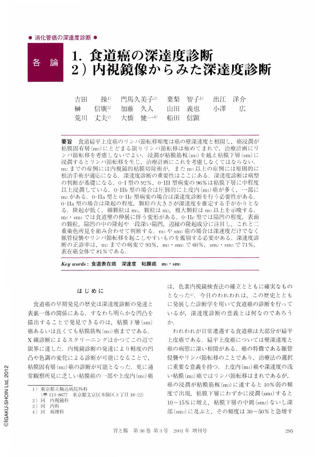
要旨 食道扁平上皮癌のリンパ節転移頻度は癌の壁深達度と相関し,癌浸潤が粘膜固有層(m2)にとどまる限りリンパ節転移は極めてまれで,治療計画にリンパ節転移を考慮しないでよい.浸潤が粘膜筋板(m3)を越え粘膜下層(sm)に浸潤するとリンパ節転移を生じ,治療計画にこれを考慮しなくてはならない.m2までの症例には内視鏡的粘膜切除術が,またm3以上の症例には原則的に根治手術が適応になる.深達度診断の重要性はここにある.深達度診断は病型の判断が基礎になる.0-Ⅰ型の92%,0-Ⅲ型病変の96%は粘膜下層に中程度以上浸潤している.0-Ⅱb型の場合は圧倒的に上皮内(m1)癌が多く,一部にm2がある.0-Ⅱa型と0-Ⅱc型病変の場合は深達度診断を行う必要性がある.0-Ⅱa型の場合は隆起の程度,顆粒の大きさが深達度を推定する手がかりとなる.隆起が低く,細顆粒はm1,顆粒はm2,粗大顆粒はm3以上を示唆する.m3・sm1では食道壁の伸展に伴う変形がある.0-Ⅱc型では陥凹の程度,表面の顆粒,陥凹の中の隆起や一段深い陥凹,辺縁の隆起成分に注目し,これと二重染色所見を組み合わせて判断する.m3やsm1癌の場合は深達度だけでなく脈管侵襲やリンパ節転移を起こしやすいものを鑑別する必要がある.深達度診断の正診率は,m2までの病変で93%,m3・sm1で60%,sm2・sm3で71%,表在癌全体で81%である.
Endoscopic evaluation of the depth of cancer invasion in cases with superficial esophageal cancer is important in selection of treatment. Lymph node metastasis is rare among cancers confined to the lamina propria mucosae. Therefore endoscopic mucosal resection (EMR) is recommended for them as the least invasive treatment. At the same time lymph node metastasis is frequent (44%) among submucosal cancers. Esophagectomy with lymph node dissection is recommended.
The gross classification of superficial cancer works well for differentiation of submucosal cancers from mucosal cancers, such as 92% of superficial and protruded lesions (type 0-Ⅰ) and 96% of superficial and depressed (type 0-Ⅲ) lesions have submucosal invasion, while submucosal invasion is infrequent among lesions with superficial and flat type (type 0-Ⅱ: 15%). At the same time, type 0-Ⅱb lesions remained within the mucosa, but 15% of all type 0-Ⅱa and 20% of type 0-Ⅱc lesions have submucosal invasion (15% of all type 0-Ⅱ lesions). Further evaluation of type 0-Ⅱa and Ⅱc lesions should be carried out. In cases with type 0-Ⅱa, the size of the granular changes on the lesion should be analyzed. Fine granules suggest mucosal cancer and coarse granules suggest cancer invasion into the muscularis mucosae or the submucosa. In cases with type 0-Ⅱc lesions, irregularities on the lesions suggest grade of invasion, e.g. flat or fine granular changes suggest mucosal cancer, coarse granular changes suggest invasion into the muscularis mucosae and nodular changes suggest invasion into the submucosa. Endoscopic double staining can differentiate mucosal cancer as pale a blue area with or without blue dots, spots or reticulation. Nodular changes in a type 0-Ⅱc lesion and interruption of “Tataminome-folds” suggest cancer invasion into the muscularis mucosae or the submucosa. The overall accuracy rate in endoscopic estimation of cancer invasion was 81% (93% for mucosal cancer remaining within the lamina propria mucosae,60% for invasion into the muscularis mucosae or slight invasion into the submucosa and 71% for moderate or massive invasion into the submucosa).

Copyright © 2001, Igaku-Shoin Ltd. All rights reserved.


