Japanese
English
- 有料閲覧
- Abstract 文献概要
- 1ページ目 Look Inside
- 参考文献 Reference
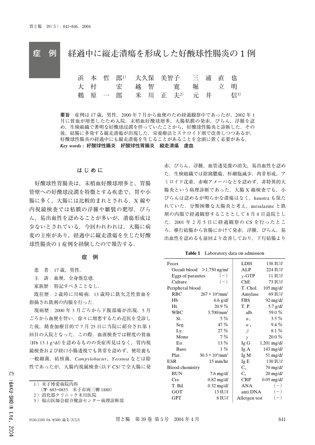
要旨 症例は17歳,男性.2000年7月から血便のため経過観察中であったが,2002年1月に貧血が増悪したため入院.末梢血好酸球増多,大腸粘膜の発赤,びらん,浮腫を認め,生検組織で著明な好酸球浸潤を伴っていたことから,好酸球性腸炎と診断した.その後,結腸に多発する縦走潰瘍が出現した.栄養療法とステロイド剤で改善しつつあるが,好酸球性腸炎の経過中にも縦走潰瘍を生じることがあることを念頭に置く必要がある.
A 17-year-old man, who had been followed up for hematochezia from2years previously, entered our hospital because of the progression of anemia. Colonoscopy showed redness, erosion and edema of the whole colonic mucosa. Biopsy specimens taken from all areas in the colon showed marked eosinophilic infiltration. The patient was diagnosed as suffering from eosinophilic enteritis. Several weeks later, follow-up colonoscopy showed longitudinal ulcer in the descending and sigmoid colon. Subsequently, he was treated with elemental diet and steroid hormone. Consequently, longitudinal ulcer became the scar while the other lesions were almost cured. We think the longitudinal ulcer in this case arose from mucosal ischemia.
1) Department of Internal Medicine, Yonago Hakuai Hospital, Yonago, Japan
2) Department of Pathology, Fukuyamashi-Ishikai General Laboratory Center, Fukuyama, Japan

Copyright © 2004, Igaku-Shoin Ltd. All rights reserved.


