Japanese
English
- 有料閲覧
- Abstract 文献概要
- 1ページ目 Look Inside
Ⅰ.はじめに
Supratentrial primitive neuroectodermal tumor(sPNET)は大脳半球に発生した未分化な神経外胚葉性細胞からなる小児脳腫瘍として1973年にHartとEarleにより提唱された6).1986年には中枢神経系に発生する中枢性PNETと末梢神経や骨・軟部組織に発生する末梢性PNETという概念が提唱され3),2000年のWHO分類でsPNETは乳幼児期に好発する稀な胎児性腫瘍に分類されている15,18).
最近,思春期に発症したsPNETの稀な1例を経験し,MRIにおける拡散強調像とFLAIR像が鑑別診断に有用であったので報告する.
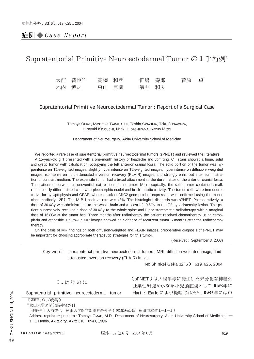
We reported a rare case of supratentorial primitive neuroectodermal tumors (sPNET) and reviewed the literature.
A 15-year-old girl presented with a one-month history of headache and vomiting. CT scans showed a huge,solid and cystic tumor with calcification,occupying the left anterior cranial fossa. The solid portion of the tumor was hypointense on T1-weighted images,slightly hyperintense on T2-weighted images,hyperintense on diffusion- weighted images,isointense on fluid-attenuated inversion recovery (FLAIR) images,and strongly enhanced after administration of contrast medium. The expansile tumor had a broad attachment to the dura matter of the anterior cranial fossa. The patient underwent an uneventful extirpation of the tumor. Microscopically,the solid tumor contained small,round poorly-differentiated cells with pleomorphic nuclei and brisk mitotic activity. The tumor cells were immunoreactive for synaptophysin and GFAP,whereas lack of MIC2 gene product expression was confirmed using the monoclonal antibody 12E7. The MIB-1-positive rate was 43%. The histological diagnosis was sPNET. Postoperatively,a dose of 30.6Gy was administrated to the whole brain and a boost of 19.6Gy to the T2-hyperintensity lesion. The patient successively received a dose of 30.4Gy to the whole spine and Linac stereotactic radiotherapy with a marginal dose of 16.8Gy at the tumor bed. Three months after radiotherapy the patient received chemotherapy using carboplatin and etoposide. Follow-up MR images showed no evidence of recurrent tumor 5 months after the radiochemotherapy.
On the basis of MR findings on both diffusion-weighted and FLAIR images,preoperative diagnosis of sPNET may be important for choosing appropriate therapeutic strategies for this tumor.

Copyright © 2004, Igaku-Shoin Ltd. All rights reserved.


