Japanese
English
- 有料閲覧
PPV購入案内
1,320円 (1,200円+税10%) こちらは、597ページから600ページの文献です。購入には医書.jp本会員登録が必要です。
会員登録完了後に改めて上記「購入サイトへ」を選択してください。
または「購入サイトへ」選択後に「購入ログイン」、「新規会員登録」の順に進んでください。
会員登録について詳しくはをご確認ください。
- Abstract 文献概要
- 1ページ目 Look Inside
Ⅰ.緒言
膨大部稜における感覚細胞を形態的および神経終末の両者からⅠ型細胞とⅡ型細胞とに分類したのはWersäll(1956)である1)。Ⅰ型細胞はフラスコ型をしており,神経終末は1コで杯状をしている。Ⅱ型細胞は円柱状をしており,神経終末は小さく,多数認められる。
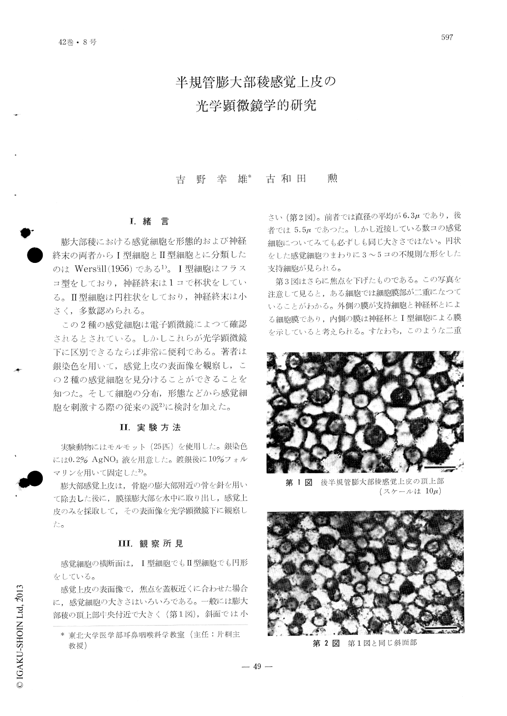
この2種の感覚細胞は電子顕微鏡によつて確認されるとされている。しかしこれらが光学顕微鏡下に区別できるならば非常に便利である。著者は銀染色を用いて,感覚上皮の表面像を観察し,この2種の感覚細胞を見分けることができることを知つた。そして細胞の分布,形態などから感覚細胞を刺激する際の従来の説2)に検討を加えた。
From the study of the sensory epithelium of the cristae ampullares stained with silver nitrate under the light microscope, two types of cells, type Ⅰ and type Ⅱ, are recognized. At the central roof region of the ampulla type Ⅰ cells predominated by 1-2 in comparison to that of the type Ⅱ while, at the region of the planum semilunatum, both types were distributed almost in equal numbers.
The type Ⅰ cells were found to be composed of 2 or 3 sets of sensory fibers.
From the standpoint of the morphology and distribution, the authors discussed and evaluated the present theory upon which the sensory cells may be stimulated.
Copyright © 1970, Igaku-Shoin Ltd. All rights reserved.