Japanese
English
- 有料閲覧
PPV購入案内
1,320円 (1,200円+税10%) こちらは、1167ページから1174ページの文献です。購入には医書.jp本会員登録が必要です。
会員登録完了後に改めて上記「購入サイトへ」を選択してください。
または「購入サイトへ」選択後に「購入ログイン」、「新規会員登録」の順に進んでください。
会員登録について詳しくはをご確認ください。
- Abstract 文献概要
- 1ページ目 Look Inside
I.はじめに
近年,脳動脈瘤の治療にあたり,脳血管造影を行わなくとも,three-dimensional CT angiogra-phy(3D-CTA)からの情報のみで手術が可能であるとの報告が多い3,11,21,29).しかし,これらの報告では脳血管造影と比較して脳動脈瘤と親血管の描出能の優秀さのみを強調した内容に終始しているように思われる.一方,術後合併症においてはpterional approachを例として取り上げても静脈系の関与が重要な要因となっている2,7,16,20,27).このようなことから,脳血管造影を行わないことで静脈系の情報が確認されないまま手術に臨んでしまうことが危惧される.3D-CTAから得られる静脈系の情報が術前検索に利用できるかは重要な問題と思われる.今回われわれは,内頸動脈周囲に介在する静脈系について3D-CTAを用い,その描出能を検討した.
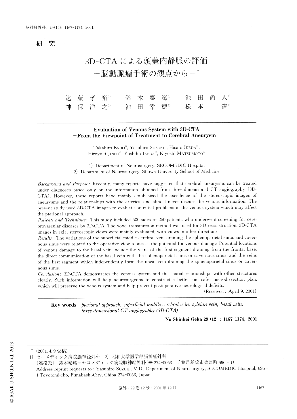
Background and Purpose: Recently, many reports have suggested that cerebral aneurysms can be treated under diagnoses based only on the information obtained from three-dimensional CT angiography (3D-CTA). However, these reports have mainly emphasized the excellence of the stereoscopic images of aneurysms and the relationships with the arteries, and almost never discuss the venous information. The present study used 3D-CTA images to evaluate potential problems in the venous system which may affect the pterional approach.
Patients and Technique: This study included 500 sides of 250 patients who underwent screening for cere-brovascular diseases by 3D-CTA. The voxel-transmission method was used for 3D reconstruction. 3D-CTA images in axial stereoscopic views were mainly evaluated, with views in other directions.
Results: The variations of the superficial middle cerebral vein draining the sphenoparietal sinus and caver-nous sinus were related to the operative view to assess the potential for venous damage. Potential locations of venous damage to the basal vein include the veins of the first segment draining from the frontal base, the direct communication of the basal vein with the sphenoparietal sinus or cavernous sinus, and the veins of the first segment which independently form the uncal vein draining the sphenoparietal sinus or caver-nous sinus.
Concluston: 3D-CTA demonstrates the venous system and the spatial relationships with other structures clearly. Such information will help neurosurgeons to construct a better and safer microdissection plan, which will preserve the venous system and help prevent postoperative neurological deficits.
Copyright © 2001, Igaku-Shoin Ltd. All rights reserved.