Japanese
English
- 有料閲覧
PPV購入案内
1,320円 (1,200円+税10%) こちらは、175ページから179ページの文献です。購入には医書.jp本会員登録が必要です。
会員登録完了後に改めて上記「購入サイトへ」を選択してください。
または「購入サイトへ」選択後に「購入ログイン」、「新規会員登録」の順に進んでください。
会員登録について詳しくはをご確認ください。
- Abstract 文献概要
- 1ページ目 Look Inside
Ι.はじめに
眼窩内静脈瘤(orbital varix)は間歇性の眼球突出を特徴的臨床症状としてくる先天性の静脈のpouchであるとされている4).一方,眼窩尖端症候群は一側の動眼,滑車,外転ならびに三叉神経第1枝の症状に視神経障害を伴うものであり,進行性の黒内障と視神経萎縮を来す2).今回われわれは右眼窩尖端症候群を有した症例において,CT scanで眼窩先端部にmassを認め,手術によりorbital varixが発見され,術後より症状の著しい改善を認めた1例を経験したので報告する.
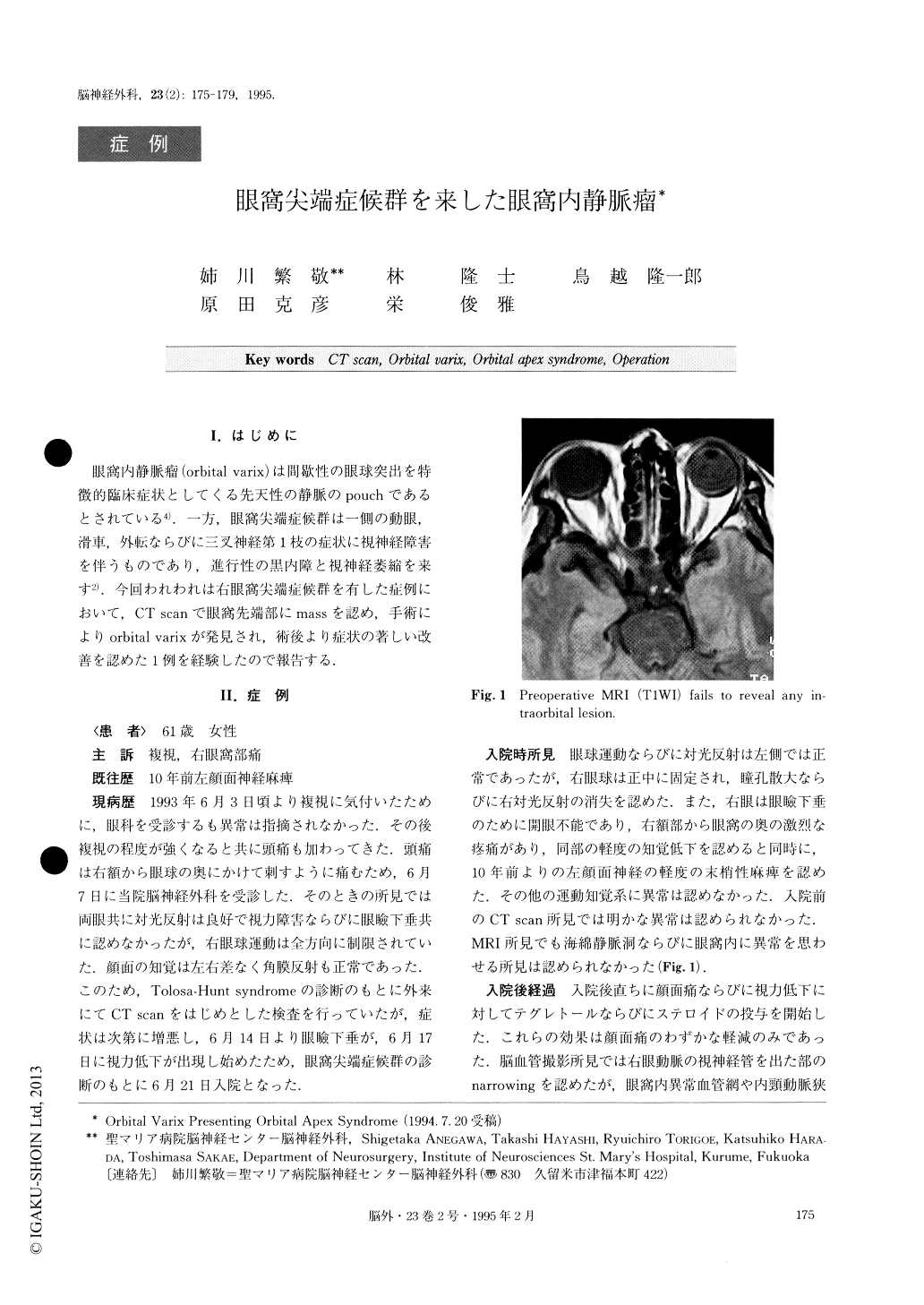
On 7 July 1993, a 61 year-old female presented a seven-day history of double vision and retro-ocular pain. Neurological examination on admission demon-strated right oculomotor palsy and pain in the rightfrontal nerve. Routine CT scan and MRI disclosed no abnormalities. Her symptoms deteriorated progressively and increased in severity, resulting in complete ptosis and visual disturbance in the right eye, for which she was hospitalized. Angiography also failed to demon-strate any lesion. Herrical CT scan demonstrated a small mass lesion at the right orbital apex.
A frontotemporal extradural approach operation was performed on July 6, 1993. The anterior clinoid process and minor wing of the sphenoid were drilled to expose the optic canal and the superior orbital fissure. When the periorbita just beside the optic sheath was opened, a large vein was observed to be impinging on the optic nerve and nerves which pass through the superior orbital fissure. This vein was carefully dissected, coagu-lated and removed. The postoperative course was un-eventful. Her severe retro-ocular pain completely dis-appeared immediately after the operation. Although complete remission of ptosis and return of eye move-ments to normal were noted, her visual acuity is at pre-sent limited to hand movement.
Orbital varix is a common cause of unilateral inter-mittent proptosis. However, there has been no report in the literature of orbital varix with orbital apex syn-drome as the initial clinical appearance. In this type of patient with orbital varix and retrobulbar hemorrhage or impairment of intraorbital nerves, surgical interven-tion using a transcranial approach to the lesion should be undertaken.
Copyright © 1995, Igaku-Shoin Ltd. All rights reserved.