Japanese
English
- 有料閲覧
PPV購入案内
1,320円 (1,200円+税10%) こちらは、485ページから490ページの文献です。購入には医書.jp本会員登録が必要です。
会員登録完了後に改めて上記「購入サイトへ」を選択してください。
または「購入サイトへ」選択後に「購入ログイン」、「新規会員登録」の順に進んでください。
会員登録について詳しくはをご確認ください。
- Abstract 文献概要
- 1ページ目 Look Inside
I.はじめに
頭蓋内出血を初発症状とする脳腫瘍は比較的まれであり,全脳腫瘍の0.8%から10.2%に認められるとされている13,16,17,19,23,26).一方,fibrosarcomaは全脳腫瘍中の0.6から1.2%に出現するとされており21,25),腫瘍出血をきたすこともあるとされているが,調べ得た限りにおいては脳内血腫により発症した本腫瘍の報告は少ない15,24).われわれは最近脳内血腫患者において,CT・脳血管撮影などの補助診断法で全く腫瘍を疑わせる所見がなく,特発性皮質下出血として手術を施行した.ところが,初回出血の約4.5ヵ月後に再度出血を繰り返し,再手術時に脳実質内より発見された腫瘍の組織学的検査によりfibrosarcomaと診断された1例を経験したので考察を加え報告する.
Abstract
A rare case of repeated intracerebral hematoma associated with an intracerebral fibrosarcoma is re-ported.
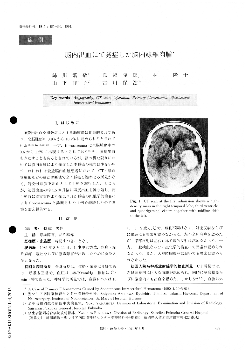
A 43-year-old man was referred to our clinic with headache and vomiting of sudden onset. On admission,he was lethargic. CT revealed a huge intracerebral hematoma in the left temporal lobe with midline shift. Angiography failed to demonstrate any evidence of an intracranial tumor. An operation was performed under the diagnosis of an idiopathic cerebral hematoma. The postoperative course was uneventful and he was dis-charged without any deficits except for a left upper quadrant homonymous hemianopia.
Four and a half months after the first operation, he was readmitted to our clinic with the same symptoms as he had at the first admission. Neuroradiological ex-amination again revealed an intracerebral hematoma in the left temporal lobe. At operation, a pinkish-gray dis-colored mass at the hematoma wall was found. An in-traoperative histological examination of the mass indi-cated a malignant tumor and the tumor was totally re-moved. However the patient did not recover from the severe neurological deficits and died 3 months after the second surgery. Histological examinations of the tumor demonstrated a typical fibrosarcoma.
Intracerebral primary fibrosarcoma with hemorrhage is quite rare. In such a case with a large hematoma, the presence of a tumor may be obscured on CT scan and angiography. Detailed observation of the hematoma wall using an operating microscope should be per-formed to allow a correct diagnosis.
Copyright © 1991, Igaku-Shoin Ltd. All rights reserved.