Japanese
English
- 有料閲覧
PPV購入案内
1,320円 (1,200円+税10%) こちらは、1181ページから1185ページの文献です。購入には医書.jp本会員登録が必要です。
会員登録完了後に改めて上記「購入サイトへ」を選択してください。
または「購入サイトへ」選択後に「購入ログイン」、「新規会員登録」の順に進んでください。
会員登録について詳しくはをご確認ください。
- Abstract 文献概要
- 1ページ目 Look Inside
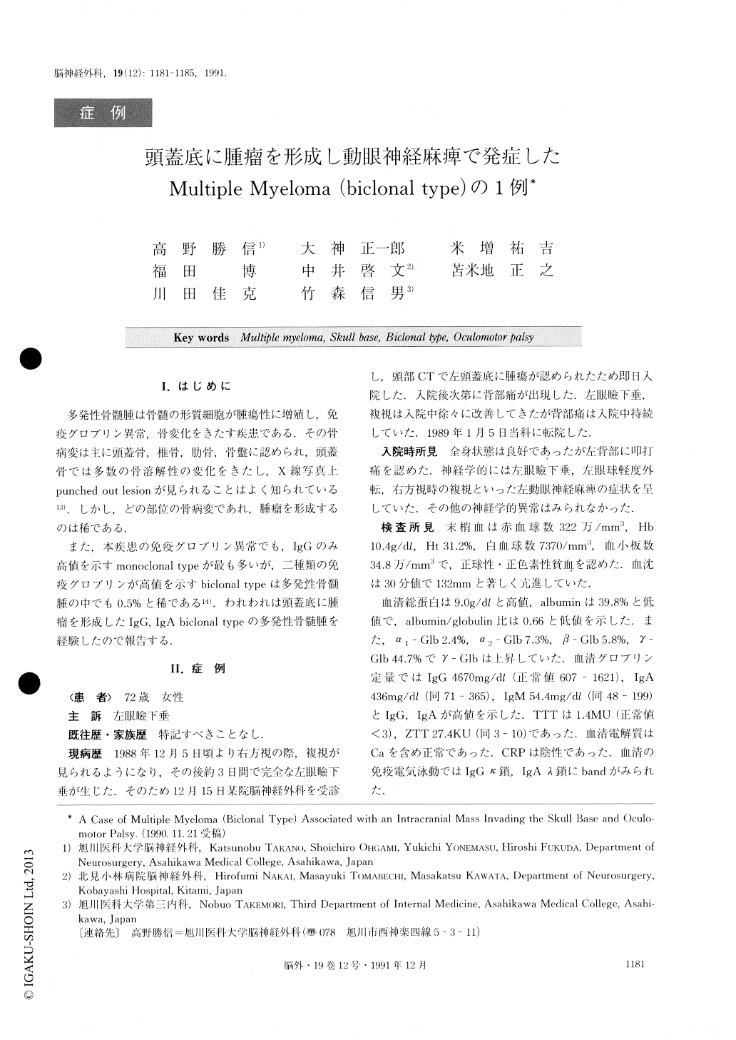
I.はじめに
多発性骨髄腫は骨髄の形質細胞が腫瘍性に増殖し,免疫グロブリン異常,骨変化をきたす疾患である.その骨病変は主に頭蓋骨,椎骨,肋骨,骨盤に認められ,頭蓋骨では多数の骨溶解性の変化をきたし,X線写真上punched out lesionが見られることはよく知られている13).しかし,どの部位の骨病変であれ,腫瘤を形成するのは稀である.
また,本疾患の免疫グロブリン異常でも,IgGのみ高値を示すmonoclonal typeが最も多いが,二種類の免疫グロブリンが高値を示すbiclonal typeは多発性骨髄腫の中でも0.5%と稀である14).われわれは頭蓋底に腫瘤を形成したIgG,IgA biclonal typeの多発性骨髄腫を経験したので報告する.
A case of multiple myeloma forming an intracranial mass which invaded the skull base was reported. A 72-year-old woman was admitted to the hospital because of left oculomotor paresis. Plain craniograms showed multiple punched out lesions. A CT scan demonstrated a mass lesion, which was homogeneously slightly en-hanced with contrast medium, in the middle cranial fos-sa. MRI, both T1 and T2 weighted images, showed an isodensity mass. In the carotid angiograms the tumor was fed by the right branches of the cavernous portionof the internal carotid artery and the maxillary artery. Laboratory data were as follows: ESR: 132mm/ 30min, serum TP: 9.0g/dl, IgG: 4670mg/dl, IgA: 430mg/d1, and urinary Bence-Jones protein was de-tected. Bone marrow biopsy of the illiac bone demon-strated myeloma cells. During hospitalization oculomo-tor paresis disappeared, and the patient was treated with intramuscular interferon_-α Multiple myeloma which invades the skull base is rare, and only 10 cases have been reported since 1977. Moreover, the biclonal type is only 0.5% of all multiple myelomas.
Copyright © 1991, Igaku-Shoin Ltd. All rights reserved.