Japanese
English
- 有料閲覧
PPV購入案内
1,320円 (1,200円+税10%) こちらは、51ページから62ページの文献です。購入には医書.jp本会員登録が必要です。
会員登録完了後に改めて上記「購入サイトへ」を選択してください。
または「購入サイトへ」選択後に「購入ログイン」、「新規会員登録」の順に進んでください。
会員登録について詳しくはをご確認ください。
- Abstract 文献概要
- 1ページ目 Look Inside
- 参考文献 Reference
Point
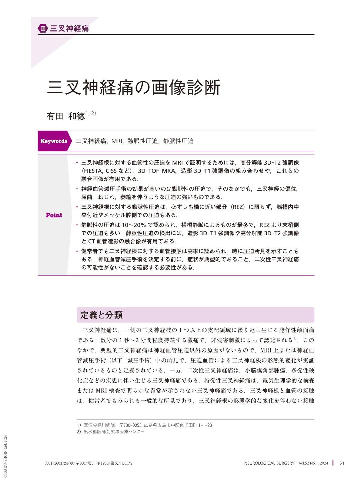
・三叉神経根に対する血管性の圧迫をMRIで証明するためには,高分解能3D-T2強調像(FIESTA, CISSなど),3D-TOF-MRA,造影3D-T1強調像の組み合わせや,これらの融合画像が有用である.
・神経血管減圧手術の効果が高いのは動脈性の圧迫で,そのなかでも,三叉神経の偏位,屈曲,ねじれ,萎縮を伴うような圧迫の強いものである.
・三叉神経根に対する動脈性圧迫は,必ずしも橋に近い部分(REZ)に限らず,脳槽内中央付近やメッケル腔側での圧迫もある.
・静脈性の圧迫は10〜20%で認められ,横橋静脈によるものが最多で,REZより末梢側での圧迫も多い.静脈性圧迫の検出には,造影3D-T1強調像や高分解能3D-T2強調像とCT血管造影の融合像が有用である.
・健常者でも三叉神経根に対する血管接触は高率に認められ,時に圧迫所見を示すこともある.神経血管減圧手術を決定する前に,症状が典型的であること,二次性三叉神経痛の可能性がないことを確認する必要性がある.
Classic trigeminal neuralgia is mainly caused by arterial compression; most cases involve the superior cerebellar artery, followed by the anterior cerebellar, basilar, and vertebral arteries. The detection of neurovascular conflicts in trigeminal neuralgia requires special magnetic resonance imaging(MRI)modalities, including high-resolution three-dimensional(3D)-T2 sequence, 3D-time of flight angiography, 3D-T1 sequencing with gadolinium injection, and merged images of these sequences. The conflicting sites are not necessarily restricted to the root entry zone of the trigeminal nerve root and can be located more distally, proximal to the Meckel's cavum. Arterial compression and its severity, including displacement, angulation, distortion, and atrophy of the trigeminal root, are good predictors of the long-term efficacy of decompression surgery. Veins, primarily the transverse pontine vein, comprise 10%-20% of all causative vessels in trigeminal neuralgia. Gadolinium-enhanced 3D-T1 MRI and high-resolution 3D-T2 MRI merged with computed tomographic angiography are useful for detecting venous compression.
Copyright © 2024, Igaku-Shoin Ltd. All rights reserved.