Japanese
English
- 有料閲覧
PPV購入案内
1,320円 (1,200円+税10%) こちらは、634ページから643ページの文献です。購入には医書.jp本会員登録が必要です。
会員登録完了後に改めて上記「購入サイトへ」を選択してください。
または「購入サイトへ」選択後に「購入ログイン」、「新規会員登録」の順に進んでください。
会員登録について詳しくはをご確認ください。
- Abstract 文献概要
- 1ページ目 Look Inside
- 参考文献 Reference
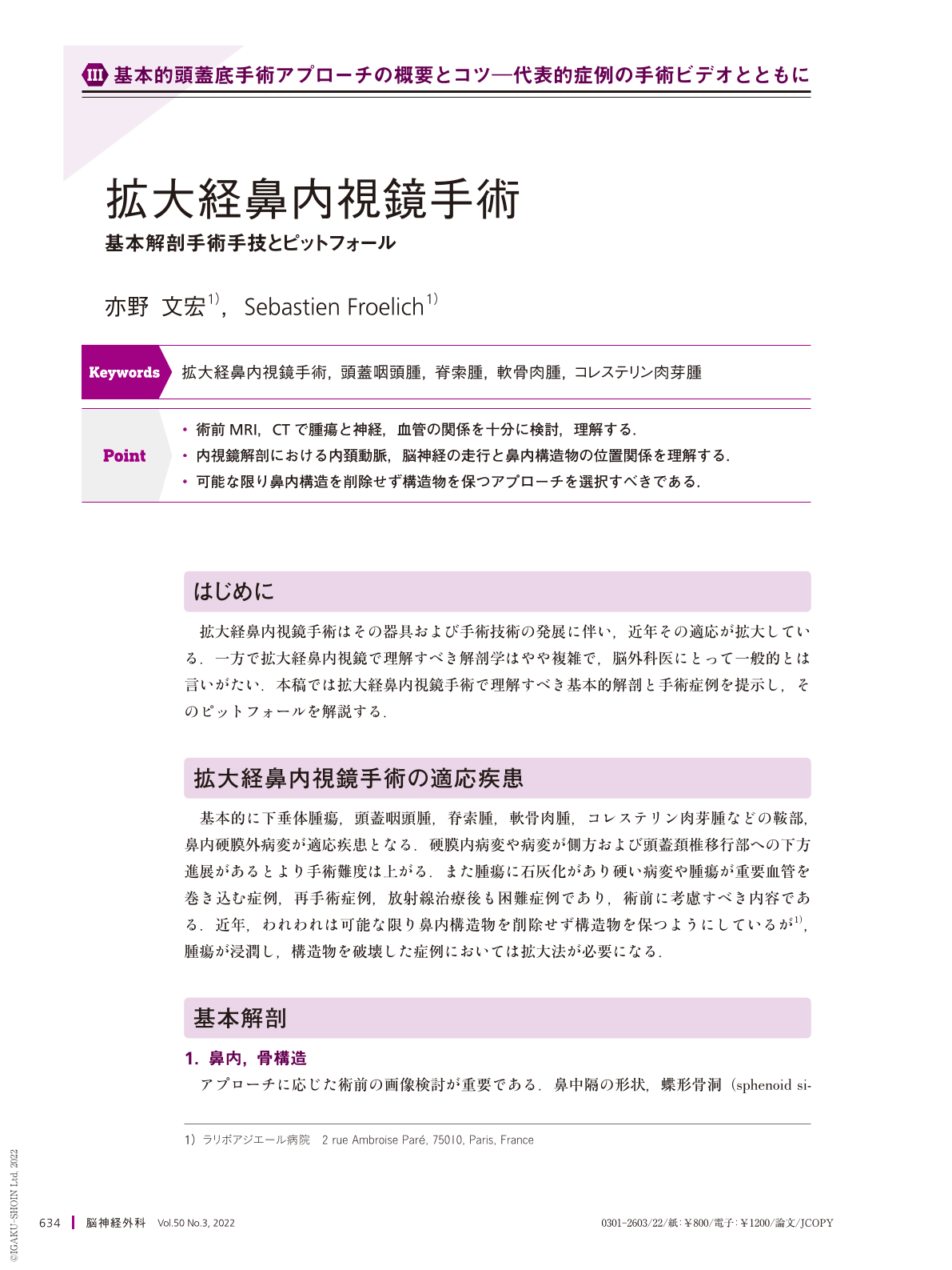
Point
・術前MRI,CTで腫瘍と神経,血管の関係を十分に検討,理解する.
・内視鏡解剖における内頚動脈,脳神経の走行と鼻内構造物の位置関係を理解する.
・可能な限り鼻内構造を削除せず構造物を保つアプローチを選択すべきである.
The extended endoscopic endonasal approach(EEA)has been expanding in recent years with the development of instruments and surgical techniques. Basically, sela, extradural, and intranasal lesions such as pituitary tumor, craniopharyngioma, chordoma, chondrosarcoma, and cholesterol granuloma are indicated. Intradural lesions or lesions that extend laterally or downward to the craniocervical junction are more difficult to operate. In addition, cases of hard tumor with calcification of the tumor, cases in which the tumor involves important blood vessels, re-operative cases, and cases after radiotherapy are also difficult cases and should be considered preoperatively. In recent years, we have been trying to keep the nasal structures as much as possible without removing nasal structure, but in cases where the tumor has invaded and destroyed the nasal structures, extended EEA is necessary. The anatomy of the extended EEA is complicated and not common among neurosurgeons. In this chapter, we present the basic anatomy and surgical cases to be understood in extended EEA and explain the pitfalls.
Copyright © 2022, Igaku-Shoin Ltd. All rights reserved.