Japanese
English
- 有料閲覧
PPV購入案内
1,320円 (1,200円+税10%) こちらは、769ページから776ページの文献です。購入には医書.jp本会員登録が必要です。
会員登録完了後に改めて上記「購入サイトへ」を選択してください。
または「購入サイトへ」選択後に「購入ログイン」、「新規会員登録」の順に進んでください。
会員登録について詳しくはをご確認ください。
- Abstract 文献概要
- 1ページ目 Look Inside
- 参考文献 Reference
Ⅰ.はじめに
類皮腫(dermoid cyst)は,胎生期遺残組織から発生する腫瘍で,皮脂腺や毛髪などの皮膚付属器を有し,鞍上部や小脳虫部,脳幹,副鼻腔,眼窩などの正中線上に好発する腫瘍である9).画像上の特徴としては,CTでは毛髪や脂肪組織による房状の低吸収域や石灰化による高吸収域を認めること,MRIではケラチンやコレステロールなどの脂肪成分を認めることとされている.しかし,腫瘍内部にはさまざまな内容物が混在するため,類皮腫の画像所見は多様であり,術前検査のみで本疾患を診断することは容易ではない10,13).今回われわれは,右前頭部に発生し,経過中に画像所見が経時的に変化した頭蓋内類皮腫の1例を経験したので,文献的考察を加えて報告する.
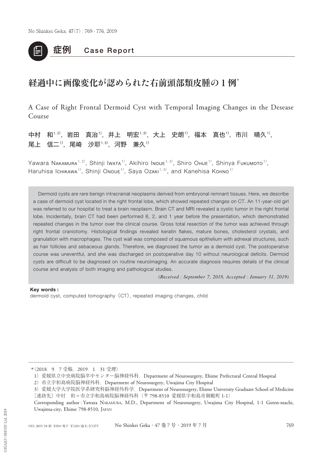
Dermoid cysts are rare benign intracranial neoplasms derived from embryonal remnant tissues. Here, we describe a case of dermoid cyst located in the right frontal lobe, which showed repeated changes on CT. An 11-year-old girl was referred to our hospital to treat a brain neoplasm. Brain CT and MRI revealed a cystic tumor in the right frontal lobe. Incidentally, brain CT had been performed 6, 2, and 1 year before the presentation, which demonstrated repeated changes in the tumor over the clinical course. Gross total resection of the tumor was achieved through right frontal craniotomy. Histological findings revealed keratin flakes, mature bones, cholesterol crystals, and granulation with macrophages. The cyst wall was composed of squamous epithelium with adnexal structures, such as hair follicles and sebaceous glands. Therefore, we diagnosed the tumor as a dermoid cyst. The postoperative course was uneventful, and she was discharged on postoperative day 10 without neurological deficits. Dermoid cysts are difficult to be diagnosed on routine neuroimaging. An accurate diagnosis requires details of the clinical course and analysis of both imaging and pathological studies.
Copyright © 2019, Igaku-Shoin Ltd. All rights reserved.