Japanese
English
- 有料閲覧
PPV購入案内
1,320円 (1,200円+税10%) こちらは、653ページから658ページの文献です。購入には医書.jp本会員登録が必要です。
会員登録完了後に改めて上記「購入サイトへ」を選択してください。
または「購入サイトへ」選択後に「購入ログイン」、「新規会員登録」の順に進んでください。
会員登録について詳しくはをご確認ください。
- Abstract 文献概要
- 1ページ目 Look Inside
- 参考文献 Reference
Ⅰ.はじめに
頭蓋外頚動脈瘤は非常に稀な疾患で,頭蓋外末梢動脈の動脈瘤全体の約0.4〜4.0%とされており1,2,6),発生部位は内頚動脈や総頚動脈に多く8,10),成因は動脈硬化や外傷によるものが多いとされる2,13).保存的に加療されることは少なく,外科的な処置が必要とされることが多いが,これまでに標準的な治療法は確立されていない.しかし,急速に増大して破裂する可能性もあるため,迅速な治療が必要となることもある.
今回われわれは,進行性に増大する拍動性頚部腫瘤で発見された頭蓋外頚動脈瘤の1例を経験したため,自験例と文献的考察を加え報告する.
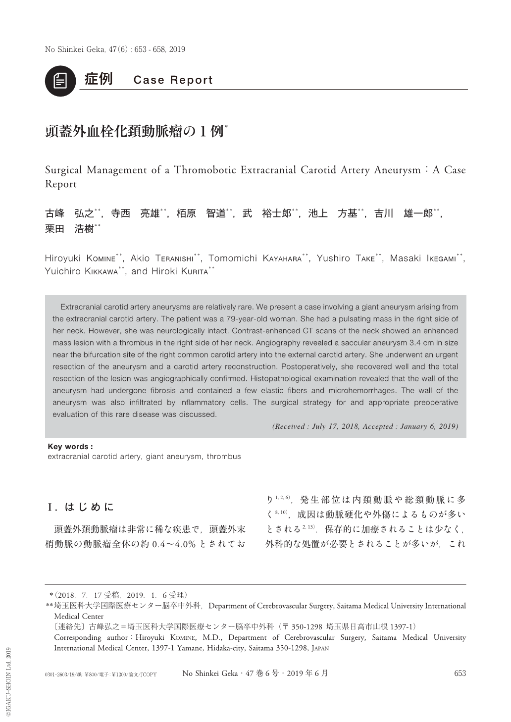
Extracranial carotid artery aneurysms are relatively rare. We present a case involving a giant aneurysm arising from the extracranial carotid artery. The patient was a 79-year-old woman. She had a pulsating mass in the right side of her neck. However, she was neurologically intact. Contrast-enhanced CT scans of the neck showed an enhanced mass lesion with a thrombus in the right side of her neck. Angiography revealed a saccular aneurysm 3.4 cm in size near the bifurcation site of the right common carotid artery into the external carotid artery. She underwent an urgent resection of the aneurysm and a carotid artery reconstruction. Postoperatively, she recovered well and the total resection of the lesion was angiographically confirmed. Histopathological examination revealed that the wall of the aneurysm had undergone fibrosis and contained a few elastic fibers and microhemorrhages. The wall of the aneurysm was also infiltrated by inflammatory cells. The surgical strategy for and appropriate preoperative evaluation of this rare disease was discussed.
Copyright © 2019, Igaku-Shoin Ltd. All rights reserved.