Japanese
English
- 有料閲覧
PPV購入案内
1,320円 (1,200円+税10%) こちらは、805ページから808ページの文献です。購入には医書.jp本会員登録が必要です。
会員登録完了後に改めて上記「購入サイトへ」を選択してください。
または「購入サイトへ」選択後に「購入ログイン」、「新規会員登録」の順に進んでください。
会員登録について詳しくはをご確認ください。
- Abstract 文献概要
- 1ページ目 Look Inside
- 参考文献 Reference
Ⅰ.はじめに
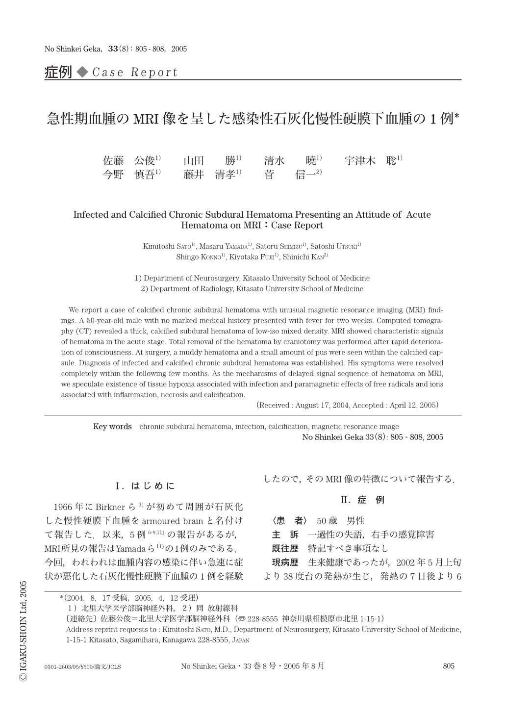
1966年にBirknerら3)が初めて周囲が石灰化した慢性硬膜下血腫をarmoured brainと名付けて報告した.以来,5例6-9,11)の報告があるが,MRI所見の報告はYamadaら11)の1例のみである.今回,われわれは血腫内容の感染に伴い急速に症状が悪化した石灰化慢性硬膜下血腫の1例を経験したので,そのMRI像の特徴について報告する.
We report a case of calcified chronic subdural hematoma with unusual magnetic resonance imaging (MRI) findings. A 50-year-old male with no marked medical history presented with fever for two weeks. Computed tomography (CT) revealed a thick,calcified subdural hematoma of low-iso mixed density. MRI showed characteristic signals of hematoma in the acute stage. Total removal of the hematoma by craniotomy was performed after rapid deterioration of consciousness. At surgery,a muddy hematoma and a small amount of pus were seen within the calcified capsule. Diagnosis of infected and calcified chronic subdural hematoma was established. His symptoms were resolved completely within the following few months. As the mechanisms of delayed signal sequence of hematoma on MRI,we speculate existence of tissue hypoxia associated with infection and paramagnetic effects of free radicals and ions associated with inflammation,necrosis and calcification.
Copyright © 2005, Igaku-Shoin Ltd. All rights reserved.