Japanese
English
- 有料閲覧
PPV購入案内
1,320円 (1,200円+税10%) こちらは、247ページから252ページの文献です。購入には医書.jp本会員登録が必要です。
会員登録完了後に改めて上記「購入サイトへ」を選択してください。
または「購入サイトへ」選択後に「購入ログイン」、「新規会員登録」の順に進んでください。
会員登録について詳しくはをご確認ください。
- Abstract 文献概要
- 1ページ目 Look Inside
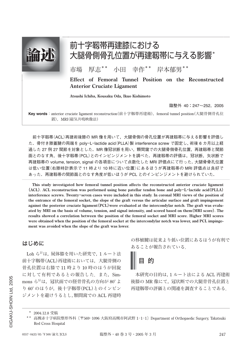
前十字靱帯(ACL)再建術後膝のMR像を用いて,大腿骨側の骨孔位置が再建靱帯に与える影響を評価した.骨付き膝蓋腱の両端をpoly-L-lactide acid(PLLA)製interference screwで固定し,術後6カ月以上経過した27例27関節を対象とした.MR像冠状断を用い,顆間窩での大腿骨側骨孔位置,再建靱帯と関節面とのなす角,後十字靱帯(PCL)とのインピンジメントを調べた.再建靱帯の評価は,冠状断,矢状断で再建靱帯のvolume,tension,signalの各項目について点数化したMRI評価点にて行った.大腿骨骨孔位置は低い位置(右膝時計表示で11時より10時に近い位置)にあるほうが再建靱帯のMRI評価点は良好であった.再建靱帯の関節面とのなす角度が低いほうがPCLとのインピンHジメントを避けられていた.
This study investigated how femoral tunnel position affects the reconstructed anterior cruciate ligament (ACL). ACL reconstruction was performed using bone patellar tendon bone and poly-L-lactide acid (PLLA) interference screws. Twenty-seven cases were included in this study. In coronal MRI views of the position of the entrance of the femoral socket, the slope of the graft versus the articular surface and graft impingement against the posterior cruciate ligament (PCL) were evaluated at the intercondylar notch. The graft was evaluated by MRI on the basis of volume, tension, and signal intensity, and scored based on them (MRI score). The results showed a correlation between the position of the femoral socket and MRI score. Higher MRI scores were obtained when the position of the femoral socket at the intercondylar notch was lower, and PCL impingement was avoided when the slope of the graft was lower.
Copyright © 2005, Igaku-Shoin Ltd. All rights reserved.