Japanese
English
- 有料閲覧
PPV購入案内
1,320円 (1,200円+税10%) こちらは、629ページから636ページの文献です。購入には医書.jp本会員登録が必要です。
会員登録完了後に改めて上記「購入サイトへ」を選択してください。
または「購入サイトへ」選択後に「購入ログイン」、「新規会員登録」の順に進んでください。
会員登録について詳しくはをご確認ください。
- Abstract 文献概要
- 1ページ目 Look Inside
- 参考文献 Reference
- サイト内被引用 Cited by
抄録
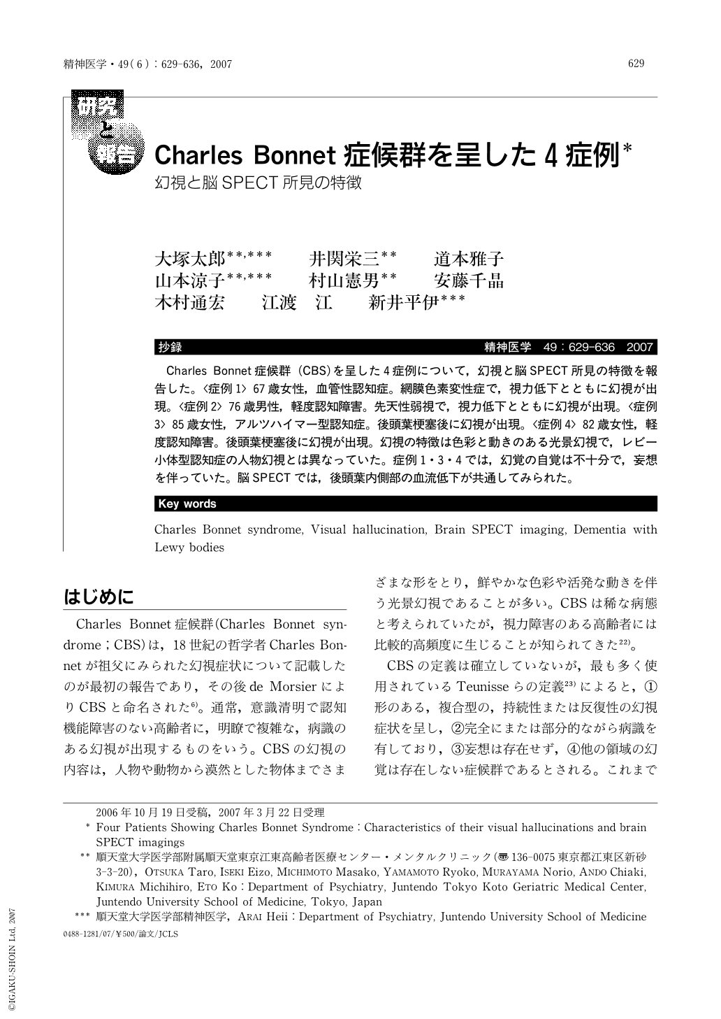
Charles Bonnet症候群 (CBS)を呈した4症例について,幻視と脳SPECT所見の特徴を報告した。〈症例1〉67歳女性,血管性認知症。網膜色素変性症で,視力低下とともに幻視が出現。〈症例2〉76歳男性,軽度認知障害。先天性弱視で,視力低下とともに幻視が出現。〈症例3〉85歳女性,アルツハイマー型認知症。後頭葉梗塞後に幻視が出現。〈症例4〉82歳女性,軽度認知障害。後頭葉梗塞後に幻視が出現。幻視の特徴は色彩と動きのある光景幻視で,レビー小体型認知症の人物幻視とは異なっていた。症例1・3・4では,幻覚の自覚は不十分で,妄想を伴っていた。脳SPECTでは,後頭葉内側部の血流低下が共通してみられた。
Charles Bonnet syndrome(CBS)means complex visual hallucinations(VH)found in elderly subjects with visual impairment and insight of VH but no delusions or other hallucinations. In this study, we reported four dementia patients with CBS who showed characteristics of VH and brain SPECT imagings.
Case 1(67-year-old female)Clinical diagnosis was vascular dementia. She developed VH, as visual impairment due to the progression of retinal degeneration. Brain MRI or SPECT showed periventricular hyperintensity or hypoperfusion in the whole occipital lobe.
Case 2(76-year-old male)Clinical diagnosis was mild cognitive impairment. He developed VH, as visual impairment due to the progression of congenital weak sight. Brain MRI or SPECT showed hippocampal atrophy or hypoperfusion in the medial occipital lobe.
Case 3(85-year-old female)Clinical diagnosis was Alzheimer-type dementia. She developed VH after a stroke. Brain MRI or SPECT showed diffuse cerebral atrophy and right occipital infarct or hypoperfusion in the parietal and medial occipital lobes, predominantly on the right side.
Case 4(82-year-old female)Clinical diagnosis was mild cognitive impairment. She developed VH after a stroke. Brain MRI or SPECT showed mild cerebral atrophy and right occipital infarct or hypoperfusion in the occipital lobe, predominantly on the right side.
Three of four cases had no sufficient insight of VH, but they had delusions. The VH of all four cases were colorfull and moving views including persons, unlike those of dementia with Lewy bodies, and their SPECT imagings shared hypoperfusion in the medial occipital lobe.
Copyright © 2007, Igaku-Shoin Ltd. All rights reserved.