Japanese
English
- 有料閲覧
PPV購入案内
1,320円 (1,200円+税10%) こちらは、1271ページから1275ページの文献です。購入には医書.jp本会員登録が必要です。
会員登録完了後に改めて上記「購入サイトへ」を選択してください。
または「購入サイトへ」選択後に「購入ログイン」、「新規会員登録」の順に進んでください。
会員登録について詳しくはをご確認ください。
- Abstract 文献概要
- 1ページ目 Look Inside
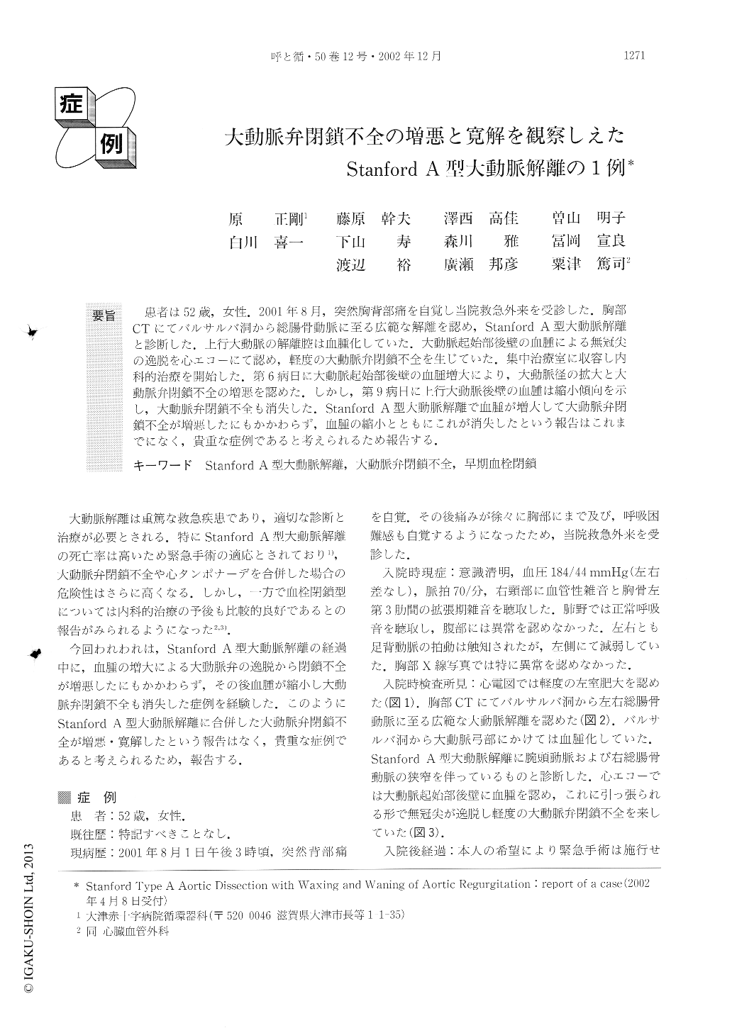
患者は52歳,女性.2001年8月,突然胸背部痛を自覚し当院救急外来を受診した.胸部CTにてバルサルバ洞から総腸骨動脈に至る広範な解離を認め,Stanford A型大動脈解離と診断した.上行大動脈の解離腔は血腫化していた.大動脈起始部後壁の血腫による無冠尖の逸脱を心エコーにて認め,軽度の大動脈弁閉鎖不全を生じていた.集中治療室に収容し内科的治療を開始した.第6病日に大動脈起始部後壁の血腫増大により,大動脈径の拡大と大動脈弁閉鎖不全の増悪を認めた.しかし,第9病日に上行大動脈後壁の血腫は縮小傾向を示し,大動脈弁閉鎖不全も消失した.Stanford A型大動脈解離で血腫が増大して大動脈弁閉鎖不全が増悪したにもかかわらず,血腫の縮小とともにこれが消失したという報告はこれまでになく,貴重な症例であると考えられるため報告する.
We reported a case of aortic dissection in which the waxing and waning of aortic regurgitation could be observed. A 52-year-old woman was brought to the emergency room with chest and back pain. Chest CT revealed aortic dissection from the ascending aorta to the bilateral common iliac arteries, which is consistent with Stanford type A aortic dissection. The dissection in the ascending aorta had been thrombosed. Echocardio-graphy revealed mild aortic regurgitation caused by the prolapse of the aortic valve leaflet. The patient was admitted to the intensive care unit and was treated medically. On the sixth day, the diameter of the ascend-ing aorta and the maximum lumen diameter were seen to have increased, which phenomenon resulted in the worsening of aortic regurgitation. Chest CT revealed enlargement of the hematoma at the posterior wall of the ascending aorta. Fortunately, the lumen diameter of the aortic dissection and hematoma decreased on the ninth day, resulting in improvement of her aortic regur-gitation.
Copyright © 2002, Igaku-Shoin Ltd. All rights reserved.