Japanese
English
- 有料閲覧
PPV購入案内
1,320円 (1,200円+税10%) こちらは、1099ページから1106ページの文献です。購入には医書.jp本会員登録が必要です。
会員登録完了後に改めて上記「購入サイトへ」を選択してください。
または「購入サイトへ」選択後に「購入ログイン」、「新規会員登録」の順に進んでください。
会員登録について詳しくはをご確認ください。
- Abstract 文献概要
- 1ページ目 Look Inside
- 参考文献 Reference
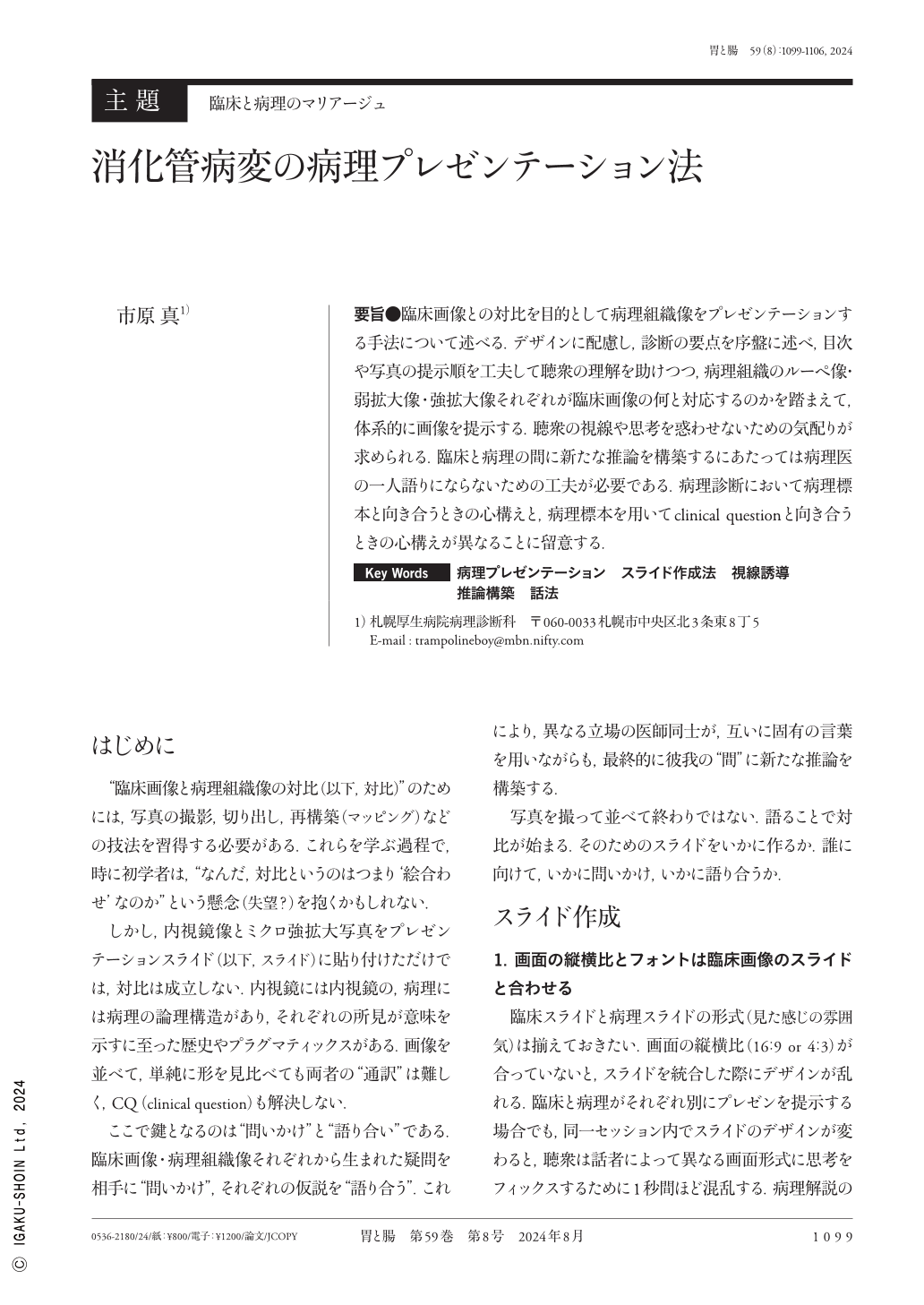
要旨●臨床画像との対比を目的として病理組織像をプレゼンテーションする手法について述べる.デザインに配慮し,診断の要点を序盤に述べ,目次や写真の提示順を工夫して聴衆の理解を助けつつ,病理組織のルーペ像・弱拡大像・強拡大像それぞれが臨床画像の何と対応するのかを踏まえて,体系的に画像を提示する.聴衆の視線や思考を惑わせないための気配りが求められる.臨床と病理の間に新たな推論を構築するにあたっては病理医の一人語りにならないための工夫が必要である.病理診断において病理標本と向き合うときの心構えと,病理標本を用いてclinical questionと向き合うときの心構えが異なることに留意する.
This report introduces a method for comparing histopathological images with clinical images. When preparing a presentation, considering its design is crucial. The main points of diagnosis should be clearly stated in the introduction. Furthermore, the table of contents and the order of presentation of photographs should be arranged thoughtfully to guide the audience's focus and enhance their understanding. Moreover, it is important to present images systematically, considering how the pathological tissue appears under various magnification levels, such as the loupe image, the weakly magnified image, and the enormously magnified image, compared with the clinical image. To avoid being the pathologist's sole narrator, it is essential to establish a new connection between clinical and pathological diagnosis. The pathologist's attitude toward pathology specimens differs for diagnosis vs. answering clinical questions.
Copyright © 2024, Igaku-Shoin Ltd. All rights reserved.