Japanese
English
- 有料閲覧
PPV購入案内
1,320円 (1,200円+税10%) こちらは、1481ページから1496ページの文献です。購入には医書.jp本会員登録が必要です。
会員登録完了後に改めて上記「購入サイトへ」を選択してください。
または「購入サイトへ」選択後に「購入ログイン」、「新規会員登録」の順に進んでください。
会員登録について詳しくはをご確認ください。
- Abstract 文献概要
- 1ページ目 Look Inside
- サイト内被引用 Cited by
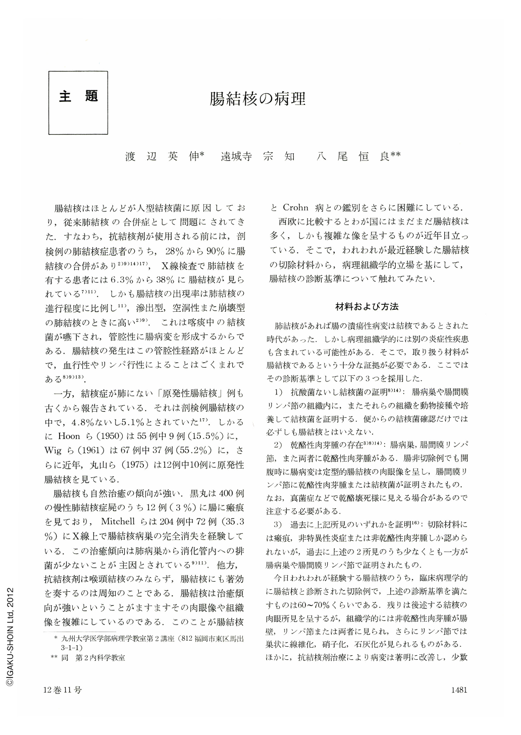
腸結核はほとんどが人型結核菌に原因しており,従来肺結核の合併症として問題にされてきた.すなわち,抗結核剤が使用される前には,剖検例の肺結核症患者のうち,28%から90%に腸結核の合併があり2)9)14)17),X線検査で肺結核を有する患者には6.3%から38%に腸結核が見られている7)11.しかも腸結核の出現率は肺結核の進行程度に比例し11),滲出型,空洞性また崩壊型の肺結核のときに高い2)9).これは喀痰中の結核菌が嚥下され,管腔性に腸病変を形成するからである.腸結核の発生はこの管腔性経路がほとんどで,血行性やリンパ行性によることはごくまれである8)9)13).
一方,結核症が肺にない「原発性腸結核」例も古くから報告されている.それは剖検例腸結核の中で,4.8%ないし5.1%とされていた17).しかるにHoonら(1950)は55例中9例(15.5%)に,Wigら(1961)は67例中37例(55.2%)に,さらに近年,丸山ら(1975)は12例中10例に原発性腸結核を見ている.
Some comparisons on histopathology and drug effects were made between 19 cases of definite intestinal tuberculosis with caseation tubercles and/or acid-fast baccilli in tissue and 10 cases of probable tuberculosis without these two findings.
The definite tuberculosis was macroscopically divided into three as follows: (1) circular ulcer which was subdivided into linear, girdle and lead-pipe forms, (2) round to oval ulcer or erosion, and (3) irregular ulcer. The first two ulcers were open and/or healed, but the last was invariably open. The ulcers were multiple and extended down to the submucosa in the majority of cases, and both open and healed ones existed together even in the non-medicated cases, and ulcers were homogenously healed usually in the medicated cases. The tubercles in the non-medicated cases were caseating and/or non-caseating, large and confluent, and, in addition, small or occasionally atrophic when observed in the intestinal wall as well as in the lymph node. The tubercles in the intestinal wall of the medicated cases were generally atrophic because of a decrease in number and size of epithelioid cells, but contained giant cells and lymphocytes which formed irregular rim in general. Especially, foreign body giant cells were prominent in the atrophic tubercles. At last these tubercles in the wall disappeared completely in the vast majority of cases. The tubercles in the lymph node often showed fibrosis, hyalinization, paralymoid deposits, and atrophy devoid of sclerotic change.
In the cases of probable tuberculosis, the nonmedicated examples showed striking similarities in gross and microscopic findings to the non-medicated definite tuberculosis, and the medicated examples to the treated definite tuberculosis. Therefore, the probable cases of intestinal tuberculosis in our series may be dealt with equally to the definite cases, and the diagnositic criteria for intestinal tuberculosis should not be restricted to caseation granulomas and/ or acid-fast bacilli because changes by spontaneous healing or antituberculous therapy are concerned.
Copyright © 1977, Igaku-Shoin Ltd. All rights reserved.