Japanese
English
- 有料閲覧
PPV購入案内
1,320円 (1,200円+税10%) こちらは、511ページから525ページの文献です。購入には医書.jp本会員登録が必要です。
会員登録完了後に改めて上記「購入サイトへ」を選択してください。
または「購入サイトへ」選択後に「購入ログイン」、「新規会員登録」の順に進んでください。
会員登録について詳しくはをご確認ください。
- Abstract 文献概要
- 1ページ目 Look Inside
- サイト内被引用 Cited by
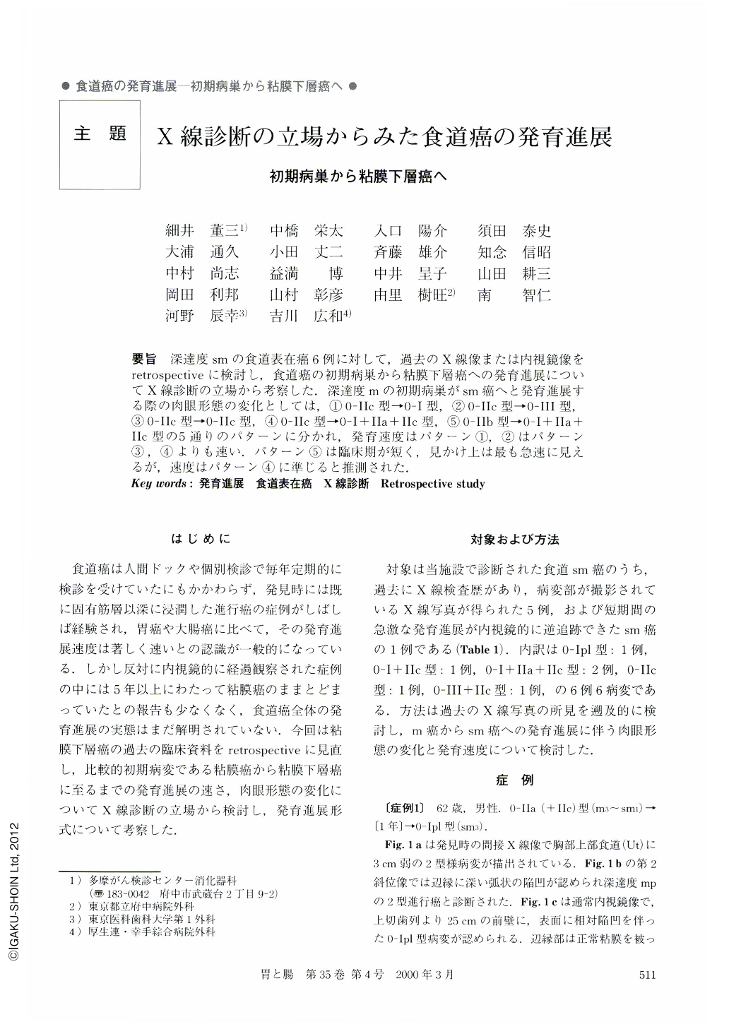
要旨 深達度smの食道表在癌6例に対して,過去のX線像または内視鏡像をretrospectiveに検討し,食道癌の初期病巣から粘膜下層癌への発育進展についてX線診断の立場から考察した.深達度mの初期病巣がsm癌へと発育進展する際の肉眼形態の変化としては,①0-IIc型→0-I型,②0-IIc型→O-III型,③0-IIc型→O-Ilc型,④0-Ilc型→O-1+IIa+IIc型,⑤0-IIb型→0-1+IIa+IIc型の5通りのパターンに分かれ,発育速度はパターン①,②はパターン③,④よりも速い.パターン⑤は臨床期が短く,見かけ上は最も急速に見えるが,速度はパターン④に準じると推測された.
Six cases of superficial esophagus cancer, all sm, were reviewed retrospectively in terms of either x-ray images or endoscopic images of the past. The growth or natural progression of esophagus cancer commencing the earliest possible stage (initial lesion) and gradually invading the submucosal layer was scrutinized from a standpoint of x-ray.
Morphological alterations where initial lesion of m develops into sm slowly and steadly were split into five patterns. ①Type 0-IIc to Type 0-I, ②Type 0-IIc to Type 0-III, ③Type 0-IIc to Type 0-IIc, ④Type 0-IIc to Type 0-I+IIa+IIc, ⑤Type 0-IIb to Type I+IIa+IIc.
Pertaining to growth velocity, patterns 1 and 2 are faster than patterns 3 and 4. Pattern 5 with the short clinical stage (period of time possible to perceive the lesion) seemingly shows extremely rapid growth, but not in fact, it conforms to pattern 4.
Copyright © 2000, Igaku-Shoin Ltd. All rights reserved.