Japanese
English
- 有料閲覧
PPV購入案内
1,320円 (1,200円+税10%) こちらは、1541ページから1547ページの文献です。購入には医書.jp本会員登録が必要です。
会員登録完了後に改めて上記「購入サイトへ」を選択してください。
または「購入サイトへ」選択後に「購入ログイン」、「新規会員登録」の順に進んでください。
会員登録について詳しくはをご確認ください。
- Abstract 文献概要
- 1ページ目 Look Inside
- サイト内被引用 Cited by
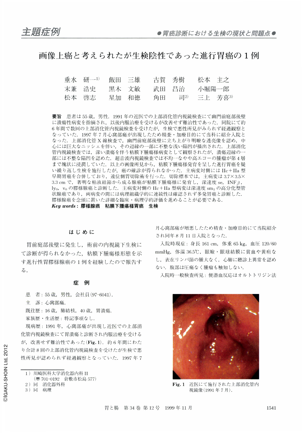
要旨 患者は55歳,男性.1991年の近医での上部消化管内視鏡検査にて幽門前庭部後壁に潰瘍性病変を指摘され,以後内服治療を受けるが改善せず難治性であった.同院にて約6年間で数回の上部消化管内視鏡検査を受けたが,生検で悪性所見がみられず経過観察となっていた.1997年7月心窩部痛が出現したため精査・加療目的にて当科に紹介入院となった.上部消化管X線検査で,幽門前庭部後壁に立ち上がり明瞭な透亮像を認め,中心には巨大なニッシェを伴い,その辺縁の一部に不整な浅い陥凹が描出された.上部消化管内視鏡検査では,深い潰瘍を伴う粘膜下腫瘍様病変として観察されたが,潰瘍辺縁の一部には不整な陥凹を認めた.超音波内視鏡検査では不均一なやや高エコーの腫瘤が第4層まで塊状に浸潤していた.以上の画像所見から,粘膜下腫瘍様発育を呈した進行胃癌を疑い繰り返し生検を施行したが,癌の確診が得られなかった.主病変対側にはⅡc+Ⅱa型早期胃癌を合併しており,遠位側胃切除術を行った.切除標本では,主病変は3.7×3.5×3.3cmで,著明な粘液結節から成る腺癌が粘膜下腫瘍様に発育し,深達度se,INFβ,ly0,v0の膠様腺癌と診断した.主病変対側のⅡc+Ⅱa型病変は深達度sm1の高分化型管状腺癌であり,両病変の間には病理組織学的に連続性は確認されず多発胃癌と診断した.膠様腺癌を念頭に置いた詳細な臨床・病理学的評価を進めることが必要である.
A 49-year-old man underwent upper endoscopy at a local clinic in 1991 because of epigastric pain. A diagnosis of gastric ulcer was made and he was treated with H2 receptor antagonist and cytoprotective agents. Repeated endoscopic examinations were performed during the next six years, with no histologic finding of carcinoma ever being found in biopsy specimens. In July 1997, he was admitted to our hospital with severe epigastric pain. Radiologic and endoscopic examinations of the stomach disclosed a submucosal tumor accompanied by a huge ulcer on the posterior wall of the antrum. Only a part of the margin of the ulcer was irregular. Endoscopic ultrasonography revealed a highechoic heterogeneous mass invading the fourth layer. Considering every finding of these imagings, advanced gastric cancer mimicking a submucosal tumor was suggested. However, carcinoma could not be confirmed by repeated histologic examinations of the biopsy specimens. Bacause another early cancer was detected on the anterior wall of the antrum, a diatal gastrectomy was performed. Macroscopic examination of the resected specimen showed an ulcerating tumor measuring 37×33 mm in size on the posterior wall and a depressed lesion measuring 20×18 mm in size on the anterior wall. Microscopic examination of the former lesion confirmed a mucinous adenocarcinoma infiltrating the serosa. Therefore, such case should be systematically evaluated by detailed clinical and careful histological examinations.
Copyright © 1999, Igaku-Shoin Ltd. All rights reserved.