Japanese
English
- 有料閲覧
- Abstract 文献概要
- 1ページ目 Look Inside
- 参考文献 Reference
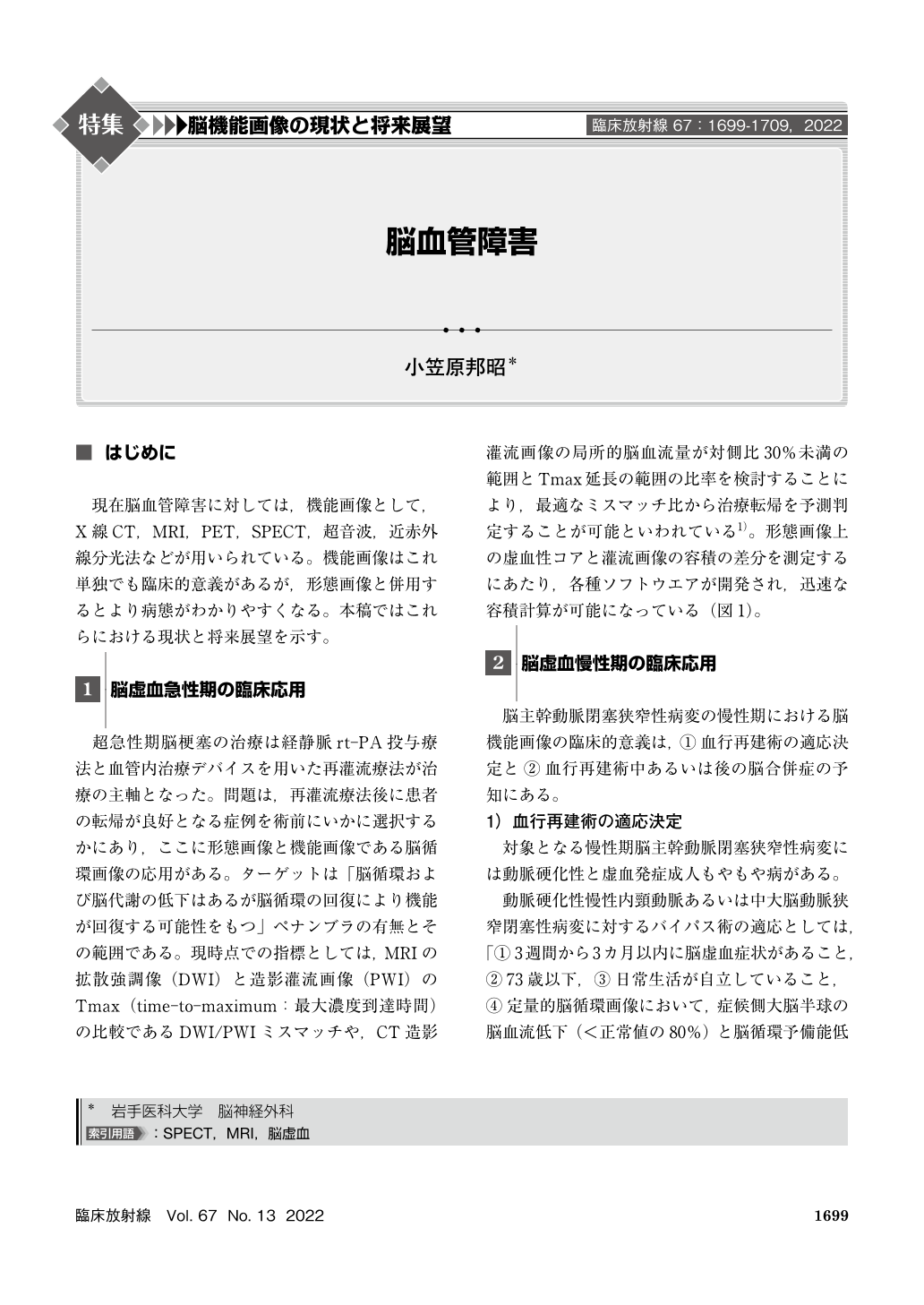
現在脳血管障害に対しては,機能画像として,X線CT,MRI,PET,SPECT,超音波,近赤外線分光法などが用いられている。機能画像はこれ単独でも臨床的意義があるが,形態画像と併用するとより病態がわかりやすくなる。本稿ではこれらにおける現状と将来展望を示す。
In reperfusion procedures including rt-PA and endovascular thrombectomy for acute ischemic stroke, ischemic penumbra is rapidly detected using diffusion-perfusion mismatch analyzed by soft wares. Brain perfusion single-photon emission computed tomography is used to determine an indication of arterial revascularization surgery for atherosclerotic cerebral main artery steno-occlusive disease or moyamoya disease or to predict cerebral hyperperfusion following carotid endarterectomy. Cerebral embolism as a complication of endarterectomy for cervical internal carotid artery stenosis is predicted by plaque imaging and computational fluid dynamics using magnetic resonance. Mechanisms of cognitive improvement after arterial revascularization surgery can be also clarified using various functional neuroimaging.

Copyright © 2022, KANEHARA SHUPPAN Co.LTD. All rights reserved.


