Japanese
English
- 有料閲覧
- Abstract 文献概要
- 1ページ目 Look Inside
- 参考文献 Reference
- サイト内被引用 Cited by
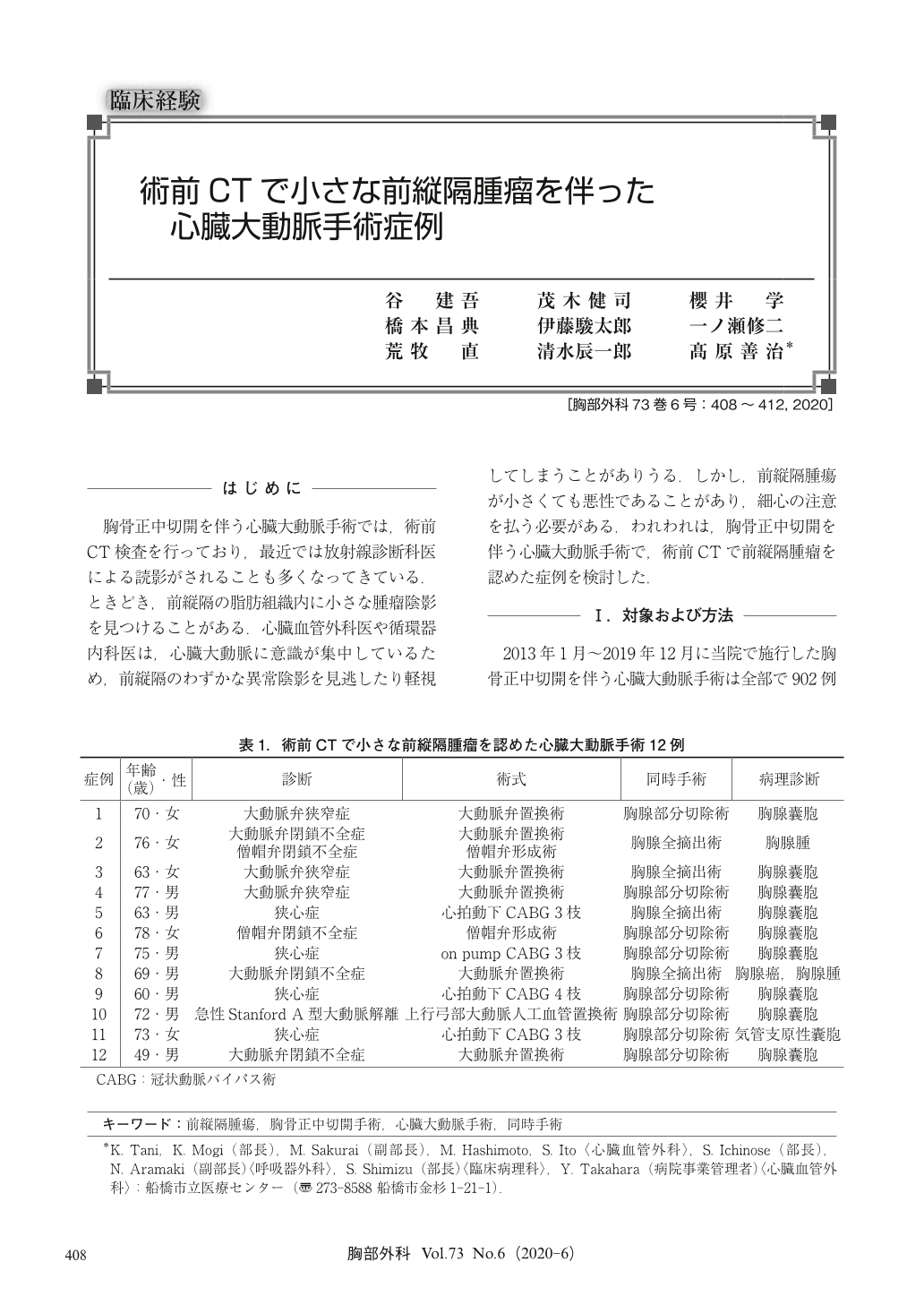
胸骨正中切開を伴う心臓大動脈手術では,術前CT検査を行っており,最近では放射線診断科医による読影がされることも多くなってきている.ときどき,前縦隔の脂肪組織内に小さな腫瘤陰影を見つけることがある.心臓血管外科医や循環器内科医は,心臓大動脈に意識が集中しているため,前縦隔のわずかな異常陰影を見逃したり軽視してしまうことがありうる.しかし,前縦隔腫瘍が小さくても悪性であることがあり,細心の注意を払う必要がある.われわれは,胸骨正中切開を伴う心臓大動脈手術で,術前CTで前縦隔腫瘤を認めた症例を検討した.
Computed tomography (CT) is indispensable for diagnostic imaging. During preoperative assessment for cardioaortic surgery, a CT examination is performed not only for diagnostic purposes but also to decide the surgical strategy. In some cases, CT demonstrates a small abnormal mass in the adipose tissue of the anterior mediastinum. Sometimes radiologists diagnose the image and send the diagnostic report to cardiologists or cardiovascular surgeons. However, they tend to limit their focus to their field of specialty. Thus, they might overlook or underestimate an abnormal mass.
Anterior mediastinal masses, though small, may include malignant tumors. Thus, we reviewed 12 cases in which anterior mediastinal masses were found on preoperative CT. Two of these patients were finally diagnosed with malignant tumors.
We should pay attention to not only cardiovascular assessment but also mediastinal masses on preoperative CT. In some cases, concomitant surgery for cardioaortic disease and an anterior mediastinal tumor is effective.

© Nankodo Co., Ltd., 2020


