Japanese
English
- 有料閲覧
- Abstract 文献概要
- 1ページ目 Look Inside
- 参考文献 Reference
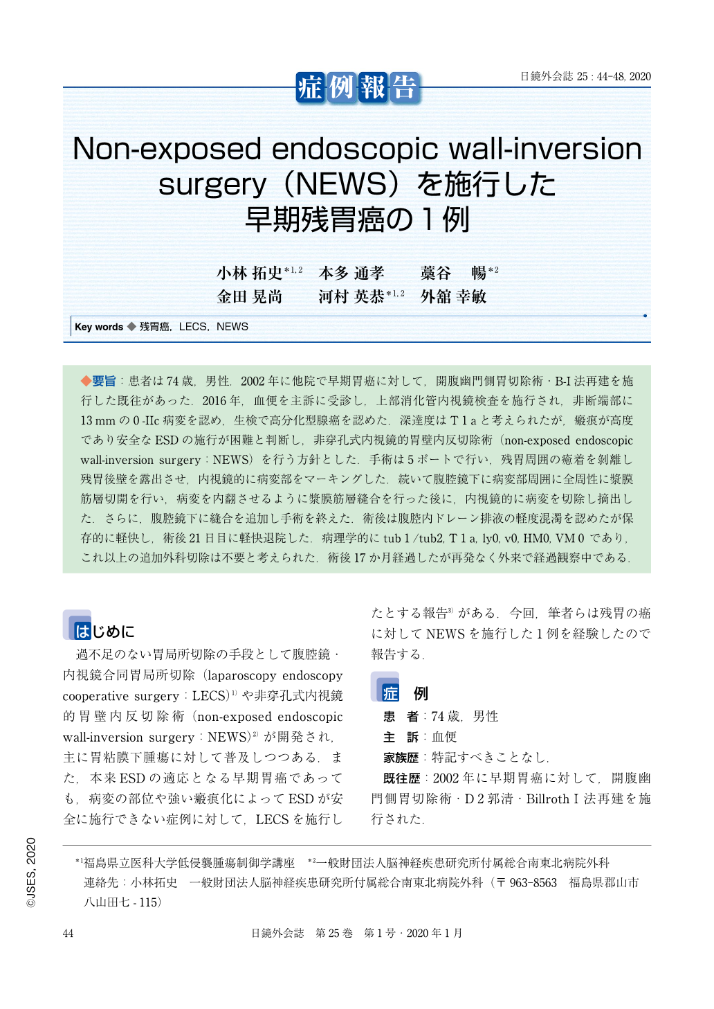
◆要旨:患者は74歳,男性.2002年に他院で早期胃癌に対して,開腹幽門側胃切除術・B-I法再建を施行した既往があった.2016年,血便を主訴に受診し,上部消化管内視鏡検査を施行され,非断端部に13mmの0-IIc病変を認め,生検で高分化型腺癌を認めた.深達度はT1aと考えられたが,瘢痕が高度であり安全なESDの施行が困難と判断し,非穿孔式内視鏡的胃壁内反切除術(non-exposed endoscopic wall-inversion surgery:NEWS)を行う方針とした.手術は5ポートで行い,残胃周囲の癒着を剝離し残胃後壁を露出させ,内視鏡的に病変部をマーキングした.続いて腹腔鏡下に病変部周囲に全周性に漿膜筋層切開を行い,病変を内翻させるように漿膜筋層縫合を行った後に,内視鏡的に病変を切除し摘出した.さらに,腹腔鏡下に縫合を追加し手術を終えた.術後は腹腔内ドレーン排液の軽度混濁を認めたが保存的に軽快し,術後21日目に軽快退院した.病理学的にtub1/tub2, T1a, ly0, v0, HM0, VM0 であり,これ以上の追加外科切除は不要と考えられた.術後17か月経過したが再発なく外来で経過観察中である.
A 74-year-old man was referred to our hospital for treatment of early remnant gastric cancer. The patient had undergone distal gastrectomy with Billroth I reconstruction 14 years ago. Gastrointestinal endoscopy showed a superficial depressed type (0-IIc) lesion located in his remnant stomach. Biopsy was performed and pathological result was well-differentiated adenocarcinoma. Non-exposed endoscopic wall-inversion surgery (NEWS) was planned because the scar around the tumor was too hard for endoscopic submucosal dissection (ESD). Adhesions around the remnant stomach was separated laparoscopically, exposing the serosal surface of the lesion. After marking the lesion under endoscopy, a circumferential seromuscular layer cutting was performed, inverting the lesion into the stomach, and suturing the seromuscular layer laparoscopically. Finally, the lesion was completely resected using ESD. There has been no evidence of recurrence or any complaints from the patient, 17 months after surgery.

Copyright © 2020, JAPAN SOCIETY FOR ENDOSCOPIC SURGERY All rights reserved.


