Japanese
English
- 有料閲覧
- Abstract 文献概要
- 1ページ目 Look Inside
- 参考文献 Reference
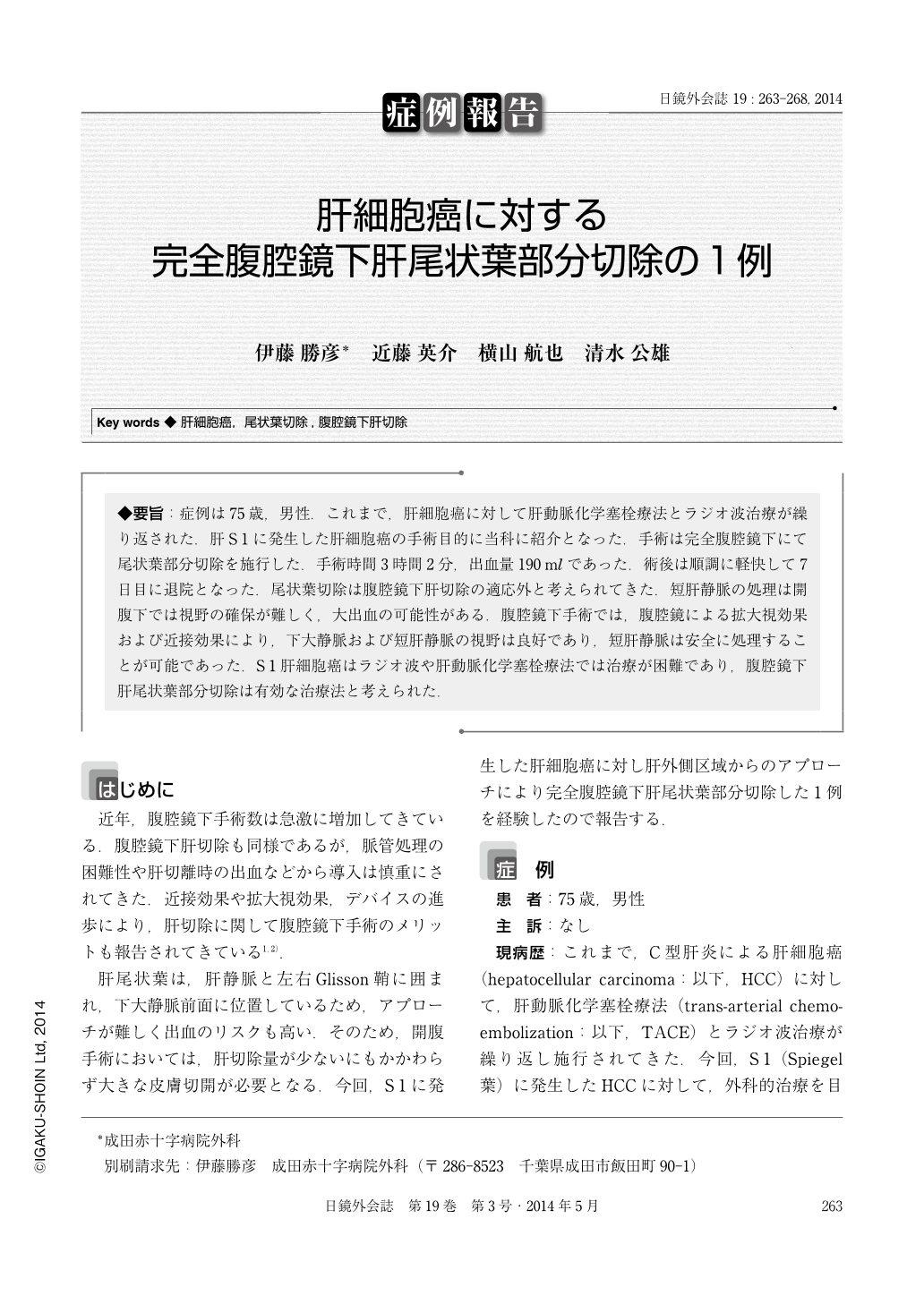
◆要旨:症例は75歳,男性.これまで,肝細胞癌に対して肝動脈化学塞栓療法とラジオ波治療が繰り返された.肝S1に発生した肝細胞癌の手術目的に当科に紹介となった.手術は完全腹腔鏡下にて尾状葉部分切除を施行した.手術時間3時間2分,出血量190mlであった.術後は順調に軽快して7日目に退院となった.尾状葉切除は腹腔鏡下肝切除の適応外と考えられてきた.短肝静脈の処理は開腹下では視野の確保が難しく,大出血の可能性がある.腹腔鏡下手術では,腹腔鏡による拡大視効果および近接効果により,下大静脈および短肝静脈の視野は良好であり,短肝静脈は安全に処理することが可能であった.S1肝細胞癌はラジオ波や肝動脈化学塞栓療法では治療が困難であり,腹腔鏡下肝尾状葉部分切除は有効な治療法と考えられた.
A 75-year-old man repeatedly underwent trans-arterial chemoembolization therapy and radiofrequency ablation therapy for hepatocellular carcinoma. He was admitted to our department because of hepatocellular carcinoma of S1 lesion. We performed pure laparoscopic partial caudate hepatectomy for the tumor. The operation time was 3 hour 2 minutes and total blood loss was 190ml. The postoperative course was uneventful and the patient was discharged on the 7 postoperative day. Laparoscopic hepatectomy for hepatocellular carcinoma arising from the caudate lobe is a surgical challenge. The deeply located tumors close to the major vessels render isolated caudate lobectomy difficult. The intrinsic illumination magnified view of the video image system offers better exposure of the lesion in the area, making resection possible even for lesion that is close to vena cava. In selected patients, laparoscopic hepatectomy for caudate lesions is feasible.

Copyright © 2014, JAPAN SOCIETY FOR ENDOSCOPIC SURGERY All rights reserved.


