Japanese
English
- 有料閲覧
- Abstract 文献概要
- 1ページ目 Look Inside
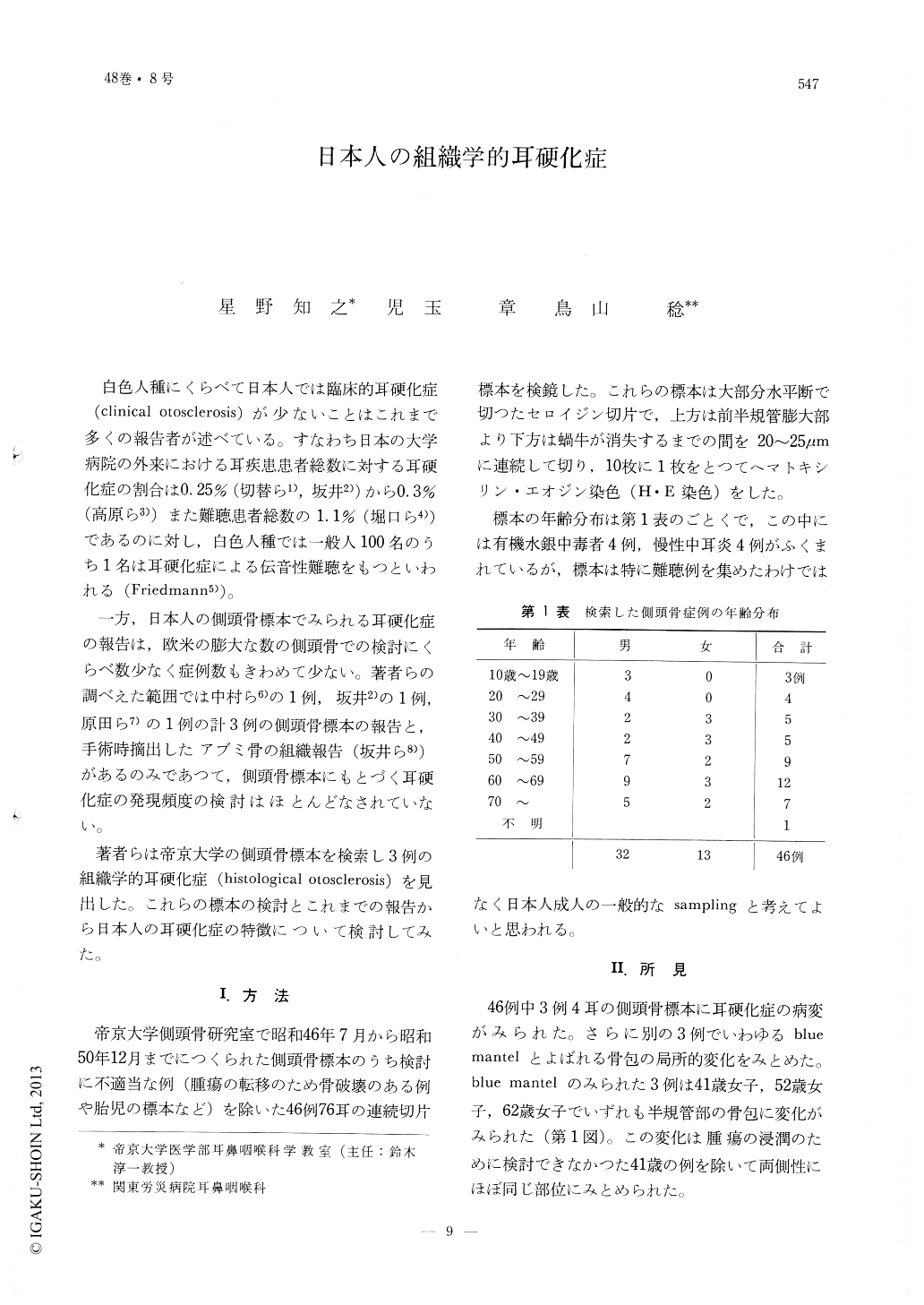
白色人種にくらべて日本人では臨床的耳硬化症(clinical otosclerosis)が少ないことはこれまで多くの報告者が述べている。すなわち日本の大学病院の外来における耳疾患患者総数に対する耳硬化症の割合は0.25%(切替ら1),坂井2))から0.3%(高原ら3))また難聴患者総数の1.1%(堀口ら4))であるのに対し,白色人種では一般人100名のうち1名は耳硬化症による伝音性難聴をもつといわれる(Friedmann5))。
一方,日本人の側頭骨標本でみられる耳硬化症の報告は,欧米の膨大な数の側頭骨での検討にくらべ数少なく症例数もきわめて少ない。著者らの調べえた範囲では中村ら6)の1例,坂井2)の1例,原田ら7)の1例の計3例の側頭骨標本の報告と,手術時摘出したアブミ骨の組織報告(坂井ら8))があるのみであつて,側頭骨標本にもとづく耳硬化症の発現頻度の検討はほとんどなされていない。
Histological evidence of otosclerosis foci was found in 4 ears, 3 individuals, out of 76 ears, 46 individuals of the Japanese temporal bone specimens studied. Bony changes were found in the left oval window area of a 64 year-old male ; in the left round window margin and the anterior wall of the internal auditory canal of a 52 year-old male ; and, in the anterior part of the right cochlea and bilateral semicircular canal regions of a 21 year-old male.
Neither diffuse nor vast invasive foci, such as frequently reported among the Caucasian temporal bones, were found in these three cases. Other reports on the Japanese temporal bone otosclerosis showed a similar restricted foci.
The authors conclude that this restricted focus development may be the underlying factor of the low incidence of clinically manifested otosclerosis among the Japanese in general.

Copyright © 1976, Igaku-Shoin Ltd. All rights reserved.


