Japanese
English
- 有料閲覧
- Abstract 文献概要
- 1ページ目 Look Inside
I.はじめに
現在,三叉神経痛に対する治療としては,mi-crovascular decompression(MVD)が優れた手術法としてほぼ確立されているが1,4,6-8),頭蓋内の三叉神経根において,臨床的に疼痛を生じている分枝の走行と,実際に圧迫されている部位の関連性については,これまで電気生理学的にほとんど検討されていない13).また,難治性の再発例や圧迫血管が見つからない症例に対しては,三叉神経根のrhizotomyが選択されることがあるが,角膜潰瘍予防のため第1枝を温存するselective rhizo-tomyの範囲を「神経根尾側の半分から3分の2」といった従来の目測による方法2,11,15)で決定した場合には,切断範囲が不正確となり,術後も神経痛が残存してしまう可能性がある.そこでわれわれは,三叉神経痛に対する手術において,三叉神経根を電気刺激し,逆行性に伝導する神経活動電位を顔面から記録することによって神経根のmappingを行い,圧迫部位における分枝の同定,およびrhizotomyの範囲決定を行ったので報告する.
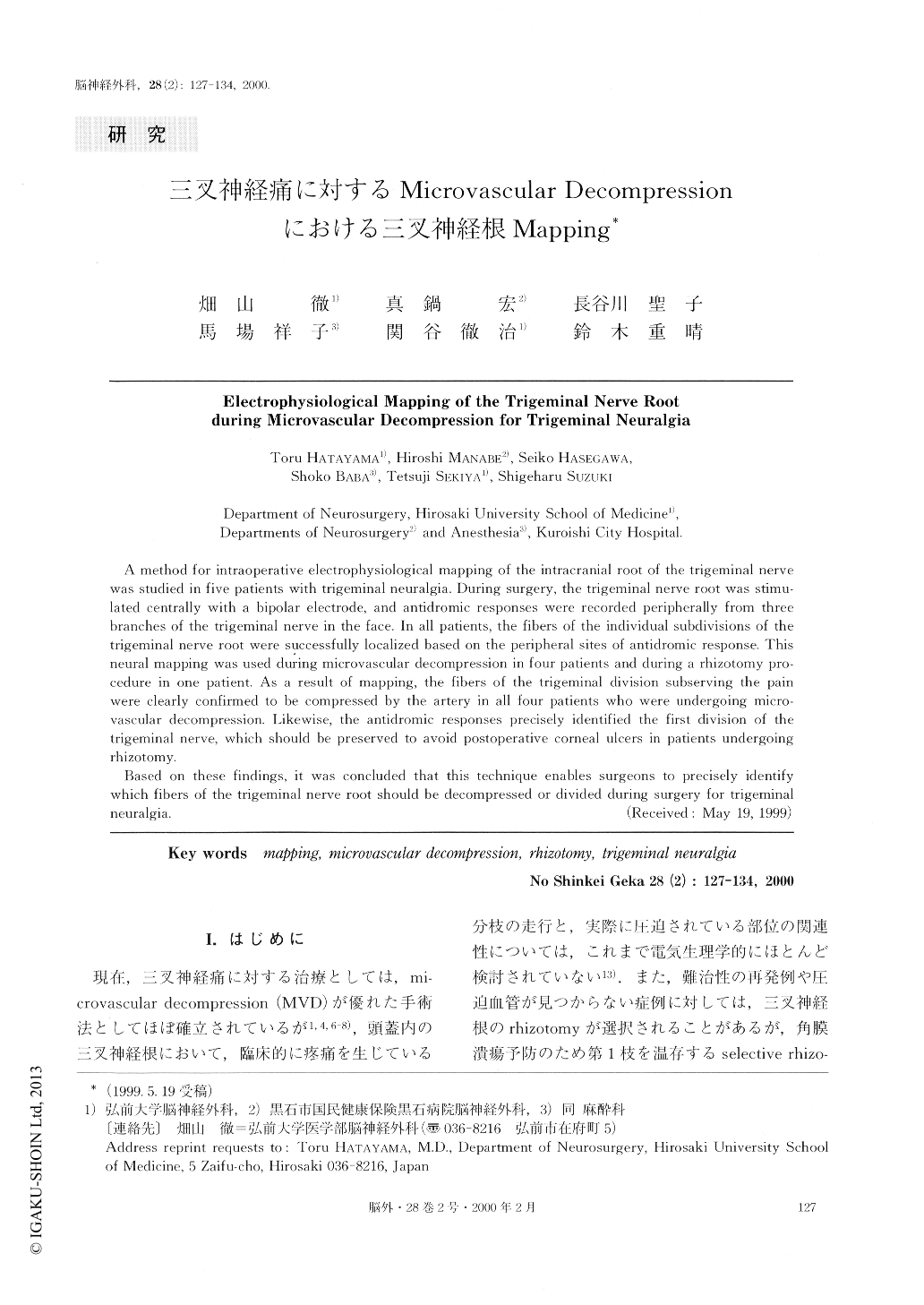
A method for intraoperative electrophysiological mapping of the intracranial root of the trigeminal nerve was studied in five patients with trigeminal neuralgia. During surgery, the trigeminal nerve root was stimu-lated centrally with a bipolar electrode, and antidromic responses were recorded peripherally from three branches of the trigeminal nerve in the face. In all patients, the fibers of the individual subdivisions of the trigeminal nerve root were successfully localized based on the peripheral sites of antidromic response. This neural mapping was used during microvascular decompression in four patients and during a rhizotomy pro-cedure in one patient. As a result of mapping, the fibers of the trigeminal division subserving the pain were clearly confirmed to be compressed by the artery in all four patients who were undergoing micro-vascular decompression. Likewise, the antidromic responses precisely identified the first division of the trigeminal nerve, which should be preserved to avoid postoperative corneal ulcers in patients undergoing rhizotomy.
Based on these findings, it was concluded that this technique enables surgeons to precisely identify which fibers of the trigeminal nerve root should be decompressed or divided during surgery for trigeminal neuralgia.

Copyright © 2000, Igaku-Shoin Ltd. All rights reserved.


