Japanese
English
- 有料閲覧
- Abstract 文献概要
- 1ページ目 Look Inside
I.はじめに
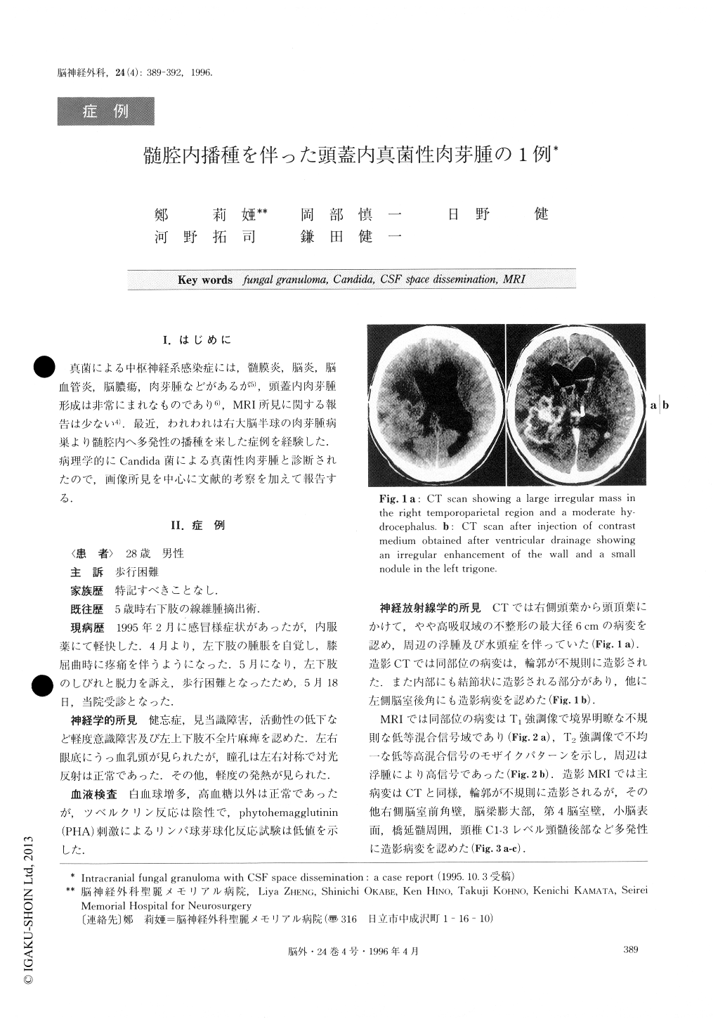
真菌による中枢神経系感染症には,髄膜炎,脳炎,脳血管炎,脳膿瘍,肉芽腫などがあるが5),頭蓋内肉芽腫形成は非常にまれなものであり6),MRI所見に関する報告は少ない4).最近,われわれは右大脳半球の肉芽腫病巣より髄腔内へ多発性の播種を来した症例を経験した.病理学的にCandida菌による真菌性肉芽腫と診断されたので,画像所見を中心に文献的考察を加えて報告する.
A 28-year-old male presented with a low grade fever, decreased activity, left hemiparesis and signs of in-tracranial hypertension. CT showed a moderate hy-drocephalus and a large irregular mass in the right tem-poroparietal region with garland-like enhancement after injection of the contract medium. These findings sug-gested a malignant brain tumor. MR images demon-strated a mass with low-iso signal intensity on T1 weighted image and low-iso-high mixed intensity on T2, which is like a mosaic pattern. Multiple cerebro-spinal fluid space seedings including the wall of the lateral ventricle, the surface of the cerebellum and pons, and the cervical spinal cord were clearly deline-ated on MR images after Gd-DTPA injection. The large mass was totally removed by craniotomy after ventricle drainage for hydrocephalus. Microscopic ex-aminations showed dense fibrous connective tissue with infiltration of Langhans' giant cells, lymphocytes and fibroblasts around the necrotic centers. These hard components may have been responsible for the low sig-nal intensity on T2-MR images. Many Candida ele-ments were clearly shown with the periodic acid Schiff stain. The diagnosis was that the lesion was an intra-parenchymal granuloma clue to Candida infection. The patient died on the 8th postoperative day because of brain stem malfunction.
Intracranial fungal infection rarely produces a granu-loma in the central nervous system. Though it is diffi-cult to diagnose a large irregular mass in the brain, MR images, especially T2 weighted images are useful for the diagnosis of fungal granuloma.

Copyright © 1996, Igaku-Shoin Ltd. All rights reserved.


