Japanese
English
- 有料閲覧
- Abstract 文献概要
- 1ページ目 Look Inside
I.はじめに
動脈瘤ネッククリッピング後の再発例に関してこれまで多数の報告例があるが紡錘状巨大動脈瘤として再発した例は非常にまれである.今回われわれは内頸動脈—後交通動脈瘤のネッククリッピングから10年経過の後,クモ膜下出血をきたした内頸動脈の紡錘状巨大動脈瘤の1例を経験したので報告する.
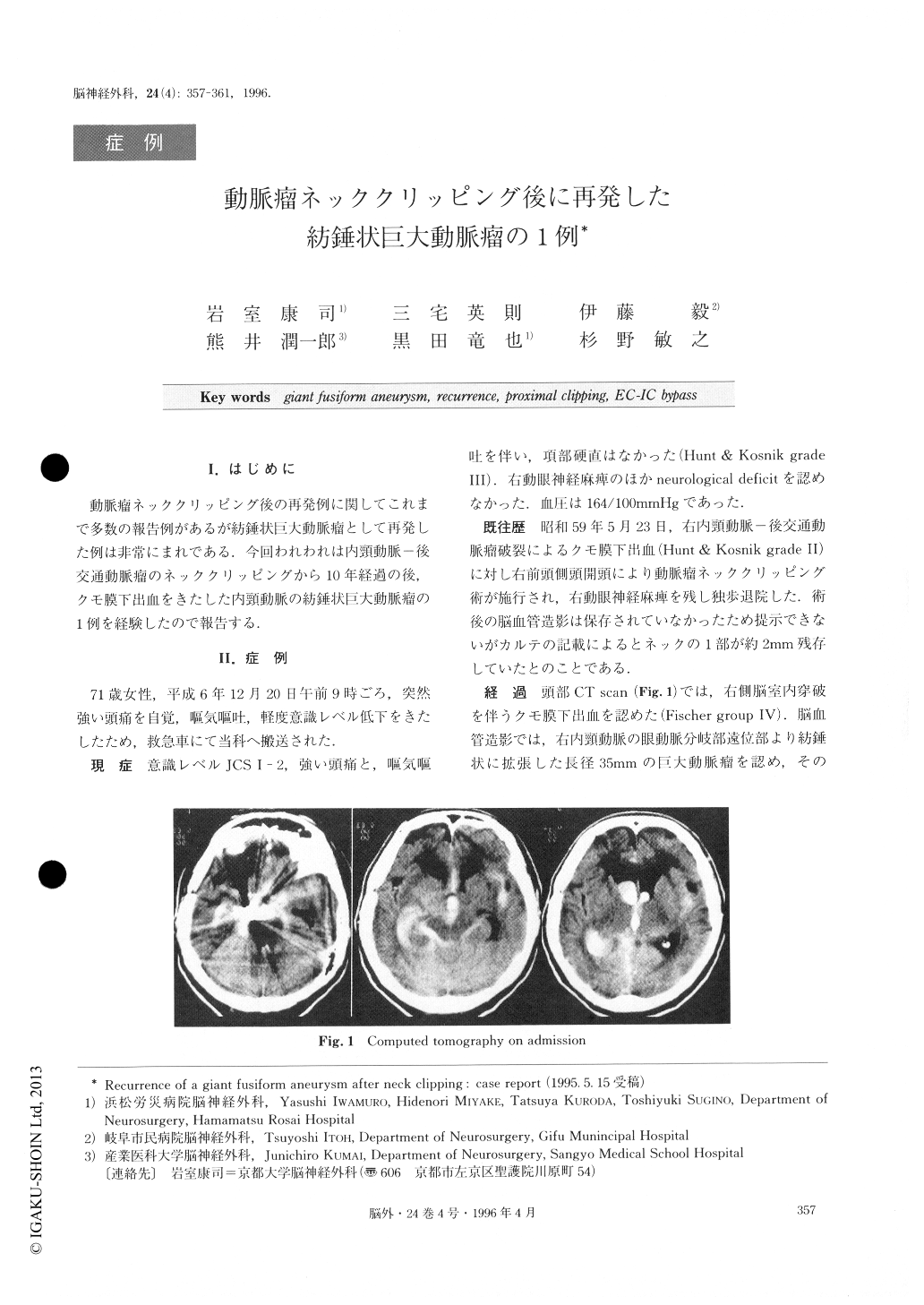
The patient was a 71-year-old female. On December 20, 1995, she suddenly developed a severe headache with vomiting and was transferred to our hospital. On admission, her conciousness level was 1-2 on the Japan Coma Scale, but there was no neurological de-ficit except for right oculomotor palsy. Computed tomograpy showed subarachnoid hemorrhage which had permeated the right lateral ventricle. On cerebral angiography, a giant fusiform aneurysm in the right in. ternal carotid artery was recognized. During the emeraencv operation, neither neck clipping nor carotid reconstruction was possible because of the tight adhe-sion of the aneurysm to the peripheral tissue. On account of this, proximal clipping of the carotid artery with external carotid-middle cerebral artery anastomosis with saphenous vein graft was selected. This patient had had an episode of subarachnoid hemorrhage owing to rupture of the right internal carotid-posterior com-municating artery aneurysm ten years earlier. At that time, the aneurysmal neck was clipped with a slight re-sidual neck and she left the hospital on foot. Five days later, when the aneurysm was found to be completely thrombosed on CT scan, antiplatelet therapy was started. Although low density areas which corre-sponded to the regions fed by the right anterior choroid-al artery were presented, re-rupture did not occur. Fol-low-up angiography showed that the aneurysm was completely thrombosed and that the right middle cere-bral and the anterior cerebral artery blood was circu-lated via the vein graft. Among recurrent cases of aneurysm after neck clipping, it is unusual for a giant fusiform aneurysm to be recognized. The growth may have been caused by sclerotic change of the arterial wall. Oculomotor palsy may have delayed the detection of the recurrence of the aneurysm. When residual neck is presented on follow-up angiography, the next angiography should be carried out within at least three years. In this case, antiplatelet therapy was effective to prevent thromboembolism from the aneurysm.

Copyright © 1996, Igaku-Shoin Ltd. All rights reserved.


