Japanese
English
- 有料閲覧
- Abstract 文献概要
- 1ページ目 Look Inside
I.はじめに
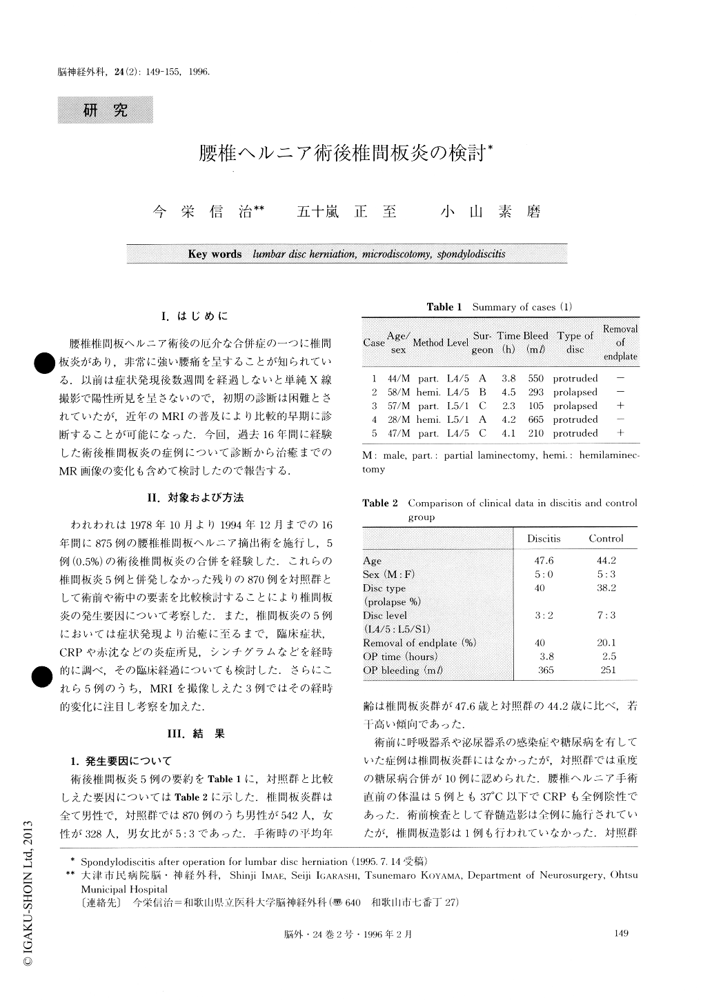
腰椎椎間板ヘルニア術後の厄介な合併症の一つに椎間板炎があり,非常に強い腰痛を呈することが知られている.以前は症状発現後数週間を経過しないと単純X線撮影で陽性所見を呈さないので,初期の診断は困難とされていたが,近年のMRIの普及により比較的早期に診断することが可能になった.今回,過去16年間に経験した術後椎間板炎の症例について診断から治癒までのMR画像の変化も含めて検討したので報告する.
Postoperative spondylodiscitis (POD) is a rare but severe complication of lumbar disc surgery. There were five patients with POD among 875 patients undergoing surgery for herniated lumbar discs, in the last 16 years.
For detecting risk factor of discitis, a POD group of 5 patients was matched to a control group of 870 pa-tients with respect to sex, age, disc type and operation. In five patients with POD follow-up evaluation of cli-nical symptoms, laboratory data were obtained, and magnetic resonance (MR) imaging was performed.
A significant difference between the POD group and the control group was confirmed in sex, operation time and the volume of bleeding during operation. However, there was no significant difference confirmed due to age, disc type, disc level and operative procedure. In the case of males or prolonged operation time or in-crease of the volume of bleeding, POD may be more frequently observed.
All five patients had a period of pain relief after their operations and then reported increasing low back pain with no focal signs. At diagnosis of POD all patients had an erythrocyte sedimentation rate (ESR) greater than 30 per hour and C-reactive protein (CRP) higher than 2.5. After treatment by antibiotics, low back pain gradually receded along with decrease of ESR and CRP. About 40 days later, these patients were almost free of back pain and ESR and CRP were within nor-mal range.
MRT1-weigted image during the acute phase demon-strated remarkably decreased signal intensity with loss of distinction between vertebral body and disc space. T2-weigted image showed increased signal intensity in the adjacent vertebral bodies and end-plates. Gadoli-nium-enhanced T1-weigted image had homogenous en-hancement of vertebral body and disc space.
During the subacute phase, however, T1-weigted im-age demonstrated moderately decreased signal intensity noted in the posteroinferior portion of the L5 vertebral body and the narrowed L5/S1 disc space. T2-weigted image showed iso signal intensity in L5 and S1 verte-bral bodies. Gadolinium-enhanced T1-weigted image had mild homogenous enhancement in the posteroinfe-rior portion of the L5 vertebral body.
Three months after treatment of POD, there was ma-jor signal change in neither T1 nor T2 weighted im-ages, though T2 showed subtle abnormalities with de-creased signal intensity adjacent to the L5/S1 disc space.
Copyright © 1996, Igaku-Shoin Ltd. All rights reserved.