Japanese
English
- 有料閲覧
- Abstract 文献概要
- 1ページ目 Look Inside
I.はじめに
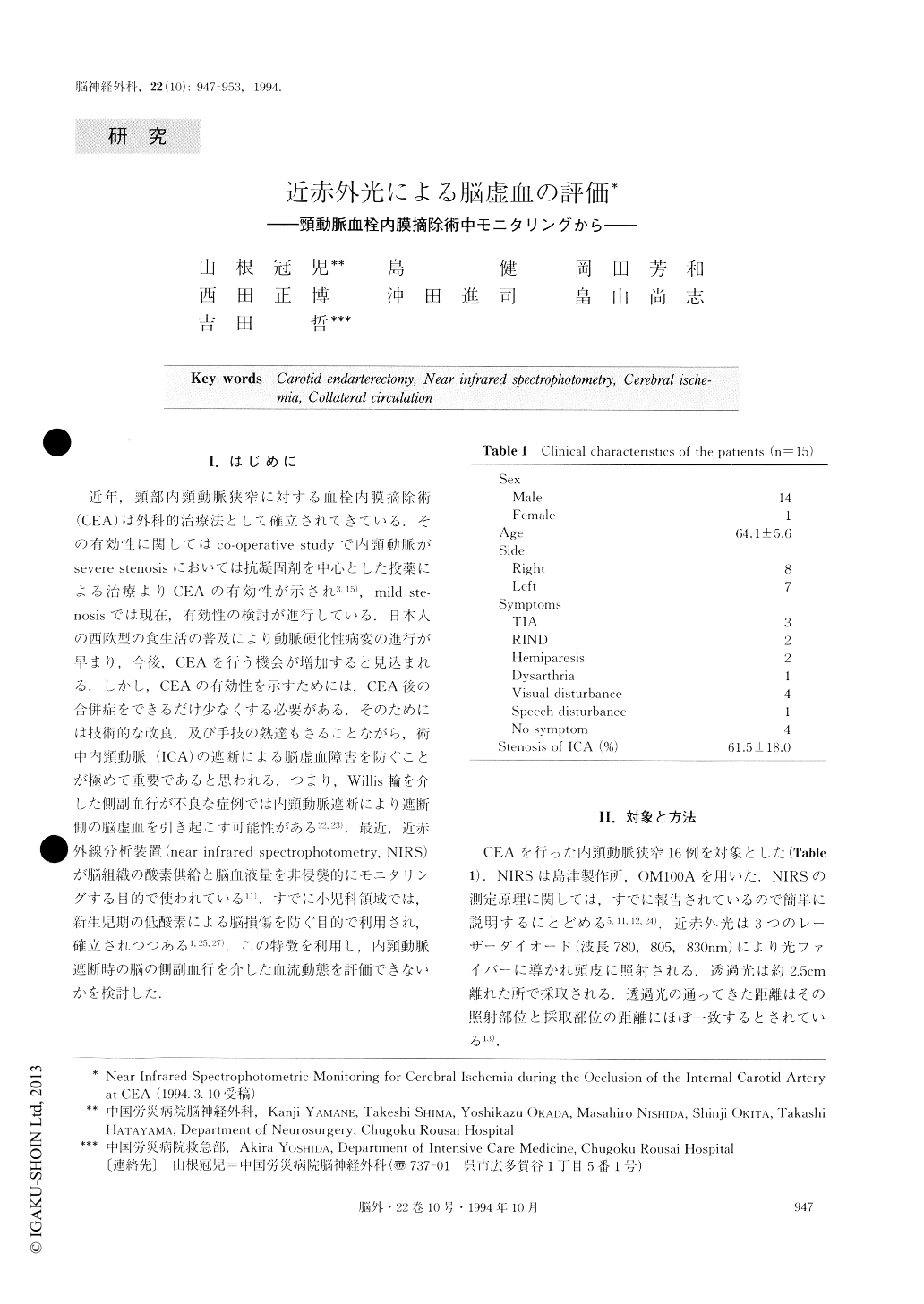
近年,頸部内頸動脈狭窄に対する血栓内膜摘除術(CEA)は外科的治療法として確立されてきている.その有効性に関してはco-operative studyで内頸動脈がsevere stenosisにおいては抗凝固剤を中心とした投薬による治療よりCEAの有効性が示され3,15),mild ste—nosisでは現在,有効性の検討が進行している.日本人の西欧型の食生活の普及により動脈硬化性病変の進行が早まり,今後,CEAを行う機会が増加すると見込まれる.しかし,CEAの有効性を示すためには,CEA後の合併症をできるだけ少なくする必要がある.そのためには技術的な改良,及び手技の熟達もさることながら,術中内頸動脈(ICA)の遮断による脳虚血障害を防ぐことが極めて重要であると思われる.つまり,Willis輪を介した側副血行が不良な症例では内頸動脈遮断により遮断側の脳虚血を引き起こす可能性がある22,23).最近,近赤外線分析装置(near infrared spectrophotometry,NIRS)が脳組織の酸素供給と脳血液量を非侵襲的にモニタリングする目的で使われている11).すでに小児科領域では,新生児期の低酸素による脳損傷を防ぐ目的で利用され,確立されつつある1,25,27).この特徴を利用し,内頸動脈遮断時の脳の側副血行を介した血流動態を評価できないかを検討した.
Near infrared spectrophotometry provides noninva-sively real-time information on cerebral oxygenation and cerebral blood volume. Using this method of spec-trophotometry we investigated the adequacy of collater-al circulation during cross-clamping of the internal carotid artery in patients who underwent carotid endar-terectomy. In 15 patients, oxy-hemoglobin, deoxy-hemoglobin and total hemoglobin were monitored con-tinuously by near infrared spectrophotometry at the ipsilateral frontal area on the operated side. Changes in these parameters following temporary cross-clamping of the internal carotid artery were evaluated. Most of the patients presented ischemic symptoms of verte-brobasilar circulation and affected upper extremities. In PTA of brachiocephalic lesions, one of the most for-midable complications is an embolism distal to the cen-tral nervous system. To prevent this complication, a vascular endoscope was used for visualization of the luminal surface of the stenotic lesions in 7 cases, and a protective balloon was used in 4 recent cases. The pro-tective balloon was used for transient occlusion of the artery to alter the flow direction so that the possible emboli might be forced to flow away to a less critical distal artery. In the distal protective balloon technique, the protective balloon was set so as to occlude the stenotic artery distally. Debris caused by PTA was aspirated and/or washed out to an extracranial artery with heparinized saline. In the proximal protective bal-loon technique, the protective balloon was set so as to occlude the stenotic artery proximally. Debris was washed out with blood flow caused by the induced steal phenomenon to an extracranial artery. In one case, in which there was stenosis in the left SA and at the left origin of the VA, kissing balloon technique was performed. Immediate post-PTA results were excellent and good in 21 cases, and poor in 3 cases including one in which the catheter could not be inserted to allow PTA to be performed. Follow-up angiography showed re-stenosis in 2 cases, and in one of them re-PTA was performed. No major complication was observed during PTA. No distal embolism was observed in any case. Vascular endoscopic observations showed the stenosed luminal surfaces before PTA were smooth and regular. No ulcer formation was observed. Therapeutic indica-tion of extracranial occlusive lesions based on our ex-perience was found in patients who had vertebrobasilar ischemic symptoms with more than 50% stenosis of VA bilaterally, or more than 50% stenosis of VA ipsilateral-ly, and aplasia or hyoplasia of VA and hypoplasia of VA at distal PICA contralaterally to the stenosis at the origin of VA. In SA stenosis, therapeutic indicaton was found in patients with ischemic symptoms of vertebro-basilar and/or upper extremity. Recent surgical techni-que for extracranial occlusive lesions is transposition of VA which has a low mortality and morbidity rate. Con-sidering the fact of invasive intervention associated with surgery and anesthesia, PTA is an alternative treat-ment. Recent reports and our experience indicate a high rate of success and rare occurrence of complications. Also safer PTA can be performed using some adjunc-tive procedures such as those mentioned in this report during PTA. The stump pressure of the internal carotid artery was mea-sured in every patient. Only the maximum decrease in oxy-hemoglobin during cross-clamping of the internal carotid artery correlated significantly with the stump pressure of the internal carotid artery. Changes in oxy-hemoglobin during cross-clamping of the internal caro-tid artery demonstrated three patterns; no change or minimally decreased (4 patients), decrease with recov-ery (4 patients), and decrease without recovery (7 patients). The stump pressure of the internal carotid artery in patients who had no recovery of their de-creased oxy-Hb was significantly lower than that in any other pattern (p<0.01, Mann-Whitney U analysis). Patients who experience decrease in oxy-Hb without recovery following cross-clamping of their internal carotid artery may have poor collateral circulation and therefore may develop cerebral ischemia.

Copyright © 1994, Igaku-Shoin Ltd. All rights reserved.


