Japanese
English
- 有料閲覧
- Abstract 文献概要
- 1ページ目 Look Inside
I.はじめに
視床は広範な大脳皮質と投射結合を持ち,第三脳室と内包の間に位置する1).周囲の複雑な神経路を反映して視床出血の症状は多彩で,進展形式と神経症状の解析から血腫部位による臨床分類が種々報告されてきたが3,12,15,18,19-21),日常診療において頻用されているものは少ない.今回われわれは中,小型の視床出血について,比較的予後と相関する簡便で実用的なCT分類を考案したので報告する.
It is not easy to predict functional outcome in pa-tients with acute-stage thalamic hemorrhage. We analys-ed 100 cases of hypertensive thalamic hemorrhage less than 4cm in diameter, and devised a practical CT clas-sification for predicting the patients' prognoses.
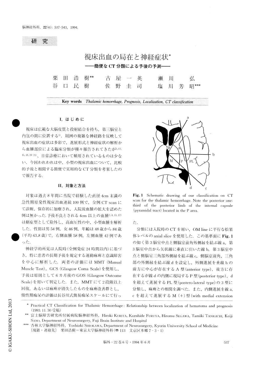
On an axial CT scan at the level of the pineal body, four lines were drawn as follows: line (a) between the lateral edge of the anterior horn and the midpoint of the third ventricle; line (b) vertical line to the sagittal line from the midpoint of the third ventricle; line (c) between the lateral edge of the trigone and the mid-point of the third ventricle; line (d) between the lateral edge of the anterior horn and the lateral edge of the tri-gone.
The location of hematoma was divided into three types according to lateral extension as follows: type A (anterior type), center of hematoma located between line (a) and line (b); type P (posterior type), center of hematoma located between line (b) and line (c), and external margin of hematoma localized medial to line (d); type PL (postero-lateral type), center of hemato-ma located between line (b) and line (c), and showing lateral extension beyond line (d). Then, the correlation between hematoma location and severity of motor pare-sis at onset and its prognosis was investigated. Severe hemiparesis (MMT: 0-2) was observed in 15.3% of patients with type A, 21.8% with type P, and 59.3% with type PL hematoma in the acute stage. Good prognosis (GOS: good recovery or moderate disability) was observed in 84.7% of patients with type A, in 70.9% with type P, but in only 12.5% with type PL hematoma (p<0.001). In our analysis, the pyra-midal tract was found to be in the posterior one-third of the posterior limb of the internal capsule, i.e. located medial to line (d) and posterior to line (b). It was con-sidered that the pyramidal tract could shift laterally without destruction in cases of P type thalamic hemor-rhage.
Hematomas were also classified as being with or without medial extension beyond both lines (a) and (c). Acute disturbance of consciousness (GCS<14) was observed in 86.4% of patients with type M+(with medial extension) and in 21.4% with type M-(without medial extension) hematoma. In addition, persistent “thalamic dementia” (Hasegawa Dementia Scale<20) limited the patient's daily life in 47.7% with type M+, but only in 8.9% with type M-(p<0.001).
This new classification does not require complicated measurements, and we can easily predict the functional outcome of motor paresis and disturbance of conscious-ness in patients with thalamic hemorrhage.

Copyright © 1994, Igaku-Shoin Ltd. All rights reserved.


