Japanese
English
- 有料閲覧
- Abstract 文献概要
- 1ページ目 Look Inside
I.はじめに
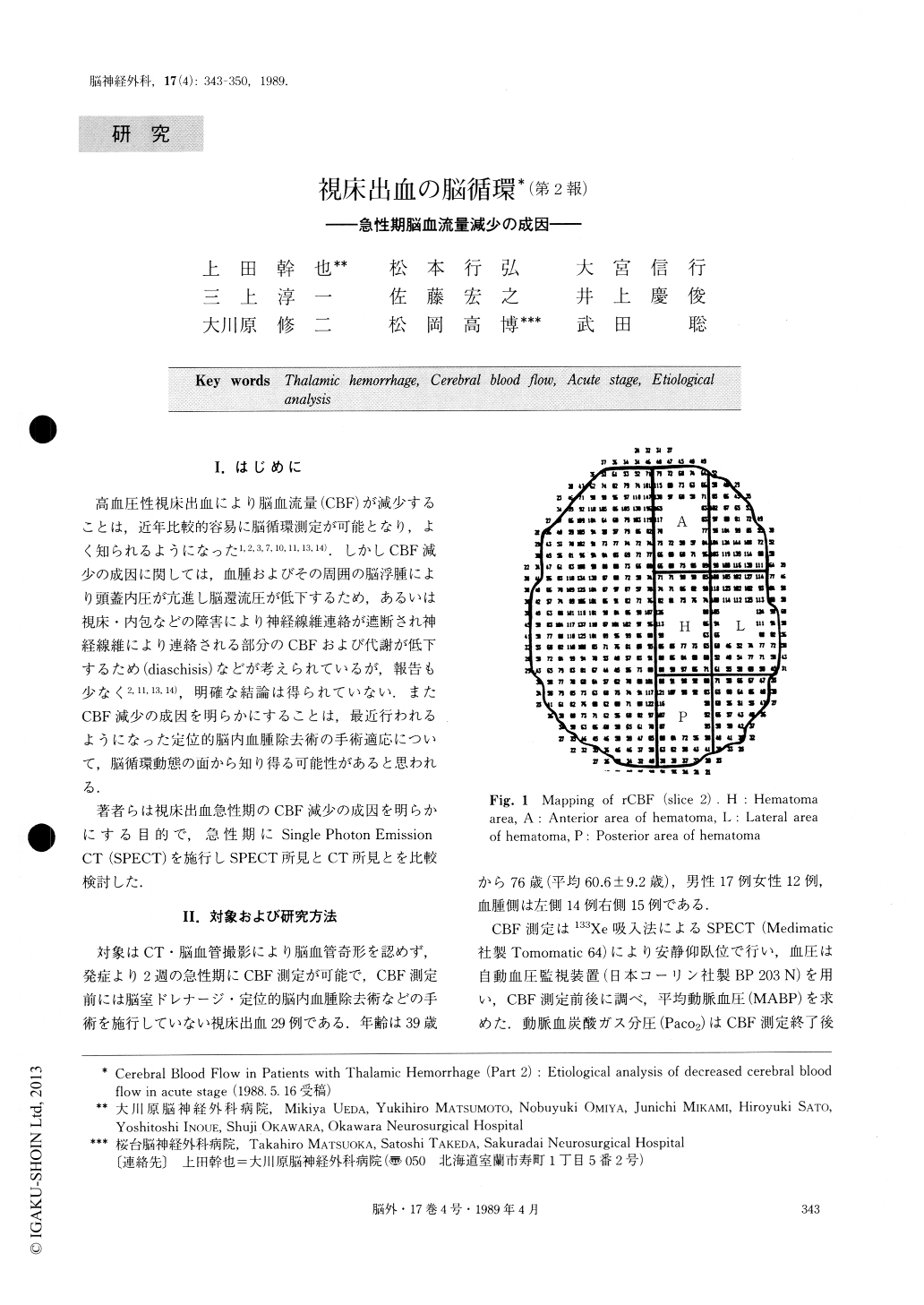
高血圧性視床出血により脳血流量(CBF)が減少することは,近年比較的容易に脳循環測定が可能となり,よく知られるようになった1,2,3,7,10,11,13,14).しかしCBF減少の成因に関しては,血腫およびその周囲の脳浮腫により頭蓋内圧が亢進し脳還流圧が低下するため,あるいは視床・内包などの障害により神経線維連絡が遮断され神経線維により連絡される部分のCBFおよび代謝が低下するため(diaschisis)などが考えられているが,報告も少なく2,11,13,14),明確な結論は得られていない.またCBF減少の成因を明らかにすることは,最近行われるようになった定位的脳内血腫除去術の手術適応について,脳循環動態の面から知り得る可能性があると思われる.
著者らは視床出血急性期のCBF減少の成因を明らかにする目的で,急性期にSingle Photon EmissionCT(SPECT)を施行しSPECT所見とCT所見とを比較検討した.
In twenty-nine patients with thalamic hemorrhage, single photon emission CT (SPECT) and CT were per-formed in the acute stage. Measurement of cerebral blood flow (CBF) was performed by the 133-Xe inhala-tion method using SPECT (Tomomatic 64) . CT find-ings such as hematoma volume, involvement of internal capsule, ventricular hematoma and topographical loca-lization of hematoma were investigated. We studied etiological analysis of decreased CBF in the acute stage.
CBF values in the group of large-volume hematoma (≧10ml) decreased moderately on the hematoma side and mildly on the nonhematoma side.

Copyright © 1989, Igaku-Shoin Ltd. All rights reserved.


