Japanese
English
- 有料閲覧
- Abstract 文献概要
- 1ページ目 Look Inside
I.はじめに
Human tail(caudal appendage)は稀な奇形であり,確実に記載されているものはこれまで数十例にすぎないようである.今回,我々はhuman tailとlipomyelo—meningoceleを有した稀な症例を経験したので若干の文献的考察を加えて報告する.
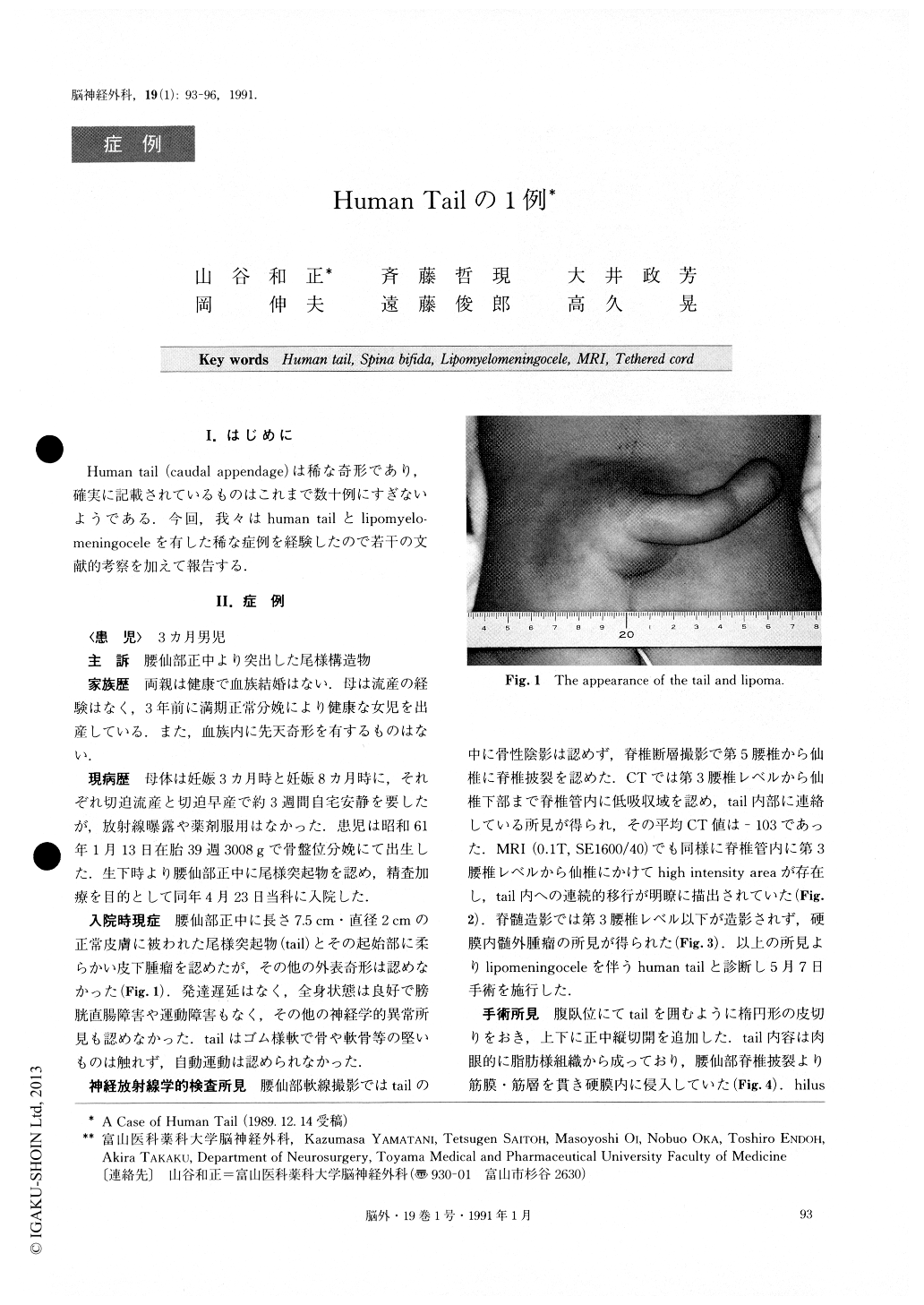
A human tail is a rare anatomical curiosity. A case of a human tail associated with lipomyelomeningocele is reported. The made subject was born, by breech deliv-ery, at the 39th-week with a 3,008 g body weight. He was admitted to our hospital because of the presence of a human tail and subcutaneous mass in the midline lumbosacral region. The tail was about 7.5 cm in length and 2 cm in diameter. It was elastic and covered by normal skin. No systemic anomaly was found. Spina bifida below L5 was revealed, and no bony shadow was found on the plain X-ray film. CT scan showed a low density area in the spinal canal between L3 and lower sacral region that extended into the tail through the spina bifida. MRI also revealed intraspinal long T2 mass which was attached to the spinal cord and ex-tended into the tail. Myelogram indicated intradural ex-tramedullary mass below the L3 level. Surgical treat-ment was performed on the 3rd month of life with a dia-gnosis of a human tail with lipomyelomeningocele. At surgery, the tail was found to consist mainly of lipoma-tous tissue which extended subcutaneously and entered the spinal canal through the spina bifida. The tail and subcutaneous lipomatous tissue were totally excised. The capsule of subcutaneous lipomatous tissue was fol-lowed circumferentially down into the spinal canal, and found to be transformed to arachnoid membrane. In-tradural lipomatous tissue was excised piece by piece, leaving only a small remnant attached to the conus medullaris to preserve sacral nerve root function. Mi-croscopic examination of the content of the tail revealed mature lipocytes, small blood vessels, myelinated nerve fibers, and longitudinally arranged striated muscle fi-bers. The patient has been free from any symptom for 2.5 years.
The authors considered that the human tail should be investigated in detail using CT or MRI because it could be combined with lipomyelomeningocele. Also, in this case, microscopical surgical treatment was necessary to prevent the development of tethered cord syndrome.

Copyright © 1991, Igaku-Shoin Ltd. All rights reserved.


