Japanese
English
- 有料閲覧
- Abstract 文献概要
- 1ページ目 Look Inside
I.はじめに
脳血管撮影で造影されないoccult AVMと呼ばれるものの中には2つの病態がある.1つは小さいかあるいは血流が緩徐であるために造影されないAVMであり,出血によって発症することが多い2,22,23).他の1つは器質化されたために造影されないthrombosed AVMと呼ばれるものであり,しばしばてんかんで発症し,手術によって診断されることが多いと言われているがその報告は少ない3-7,9-12,14,15,18,21-24).
今回われわれは,側頭葉てんかんを示したthrom—bosed AVMの症例を経験したので,放射線学的特徴とくにCTとMRI所見について文献的考察を加えて報告する.
A rare case of thrombosed AVM presenting temporal lobe epilepsy is reported. A 27-year old man was admit-ted to our hospital because of a 7-year history of tem-poral lobe epilepsy. He had also suffered from general-ized seizure since he was 6 years old.
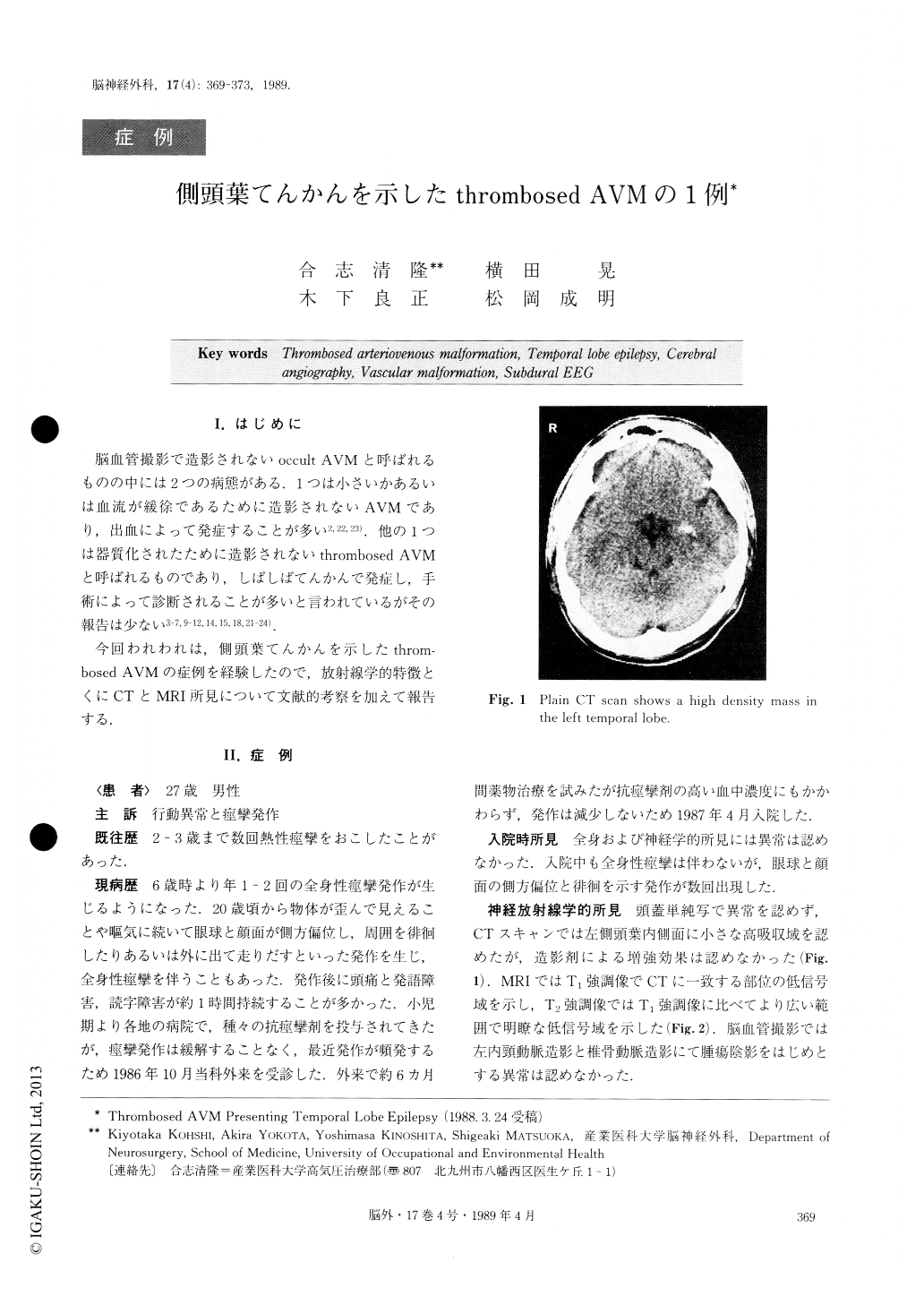
No neurological deficit was disclosed. CT scan de-monstrated a small calcified mass lesion in the left tem-poral lobe which was not enhanced by contrast study. Skull X-P and cerebral angiography were normal. Low intensity area on T1-weighted MR image corres-ponded to the high density area on CT scan.

Copyright © 1989, Igaku-Shoin Ltd. All rights reserved.


