Japanese
English
症例
MRIで髄内に病変を認めた頸髄損傷の1例—MRIの経時的所見を中心に
MRI Findings of Cervical Cord Injury: A case report of long-term follow-up
飯田 隆昭
1
,
中村 勉
1
,
飯塚 秀明
1
,
角家 暁
1
,
佐藤 秀次
2
,
伊東 正太郎
2
Takaaki IID
1
,
Tsutomu NAKAMURA
1
,
Hideaki IIZUKA
1
,
Satoru KADOYA
1
,
Shuji SATOH
2
,
Shotaro ITOH
2
1金沢医科大学脳神経外科
2金沢脳神経外科病院
1Department of Neurosurgery, Kanazawa Medica University
2Kanazawa Neurosurgical Hospital
キーワード:
MRI
,
Spinal cord injury
Keyword:
MRI
,
Spinal cord injury
pp.1501-1504
発行日 1988年12月10日
Published Date 1988/12/10
DOI https://doi.org/10.11477/mf.1436202746
- 有料閲覧
- Abstract 文献概要
- 1ページ目 Look Inside
I.はじめに
近年,脊髄髄内病変の画像診断にMRIが威力を発揮することが知られ,脊髄損傷に対してもその有用性が強調されている.すでに急性期脊髄損傷のMRI所見については実験例3,5),臨床例各々8-11)の報告がある.しかし損傷脊髄の変化をMRIで経時的に検討した報告は未だ少ない.著者らは,受傷直後より8カ月にわたってMRI所見の変化を観察できた頸髄損傷の1例を経験したので報告する.
A 63 year-old male, who sustained anterior cervical cord injury in a fall on his back during skiing, was con-secutively examined by MRI from acute, up to the chronic stage.
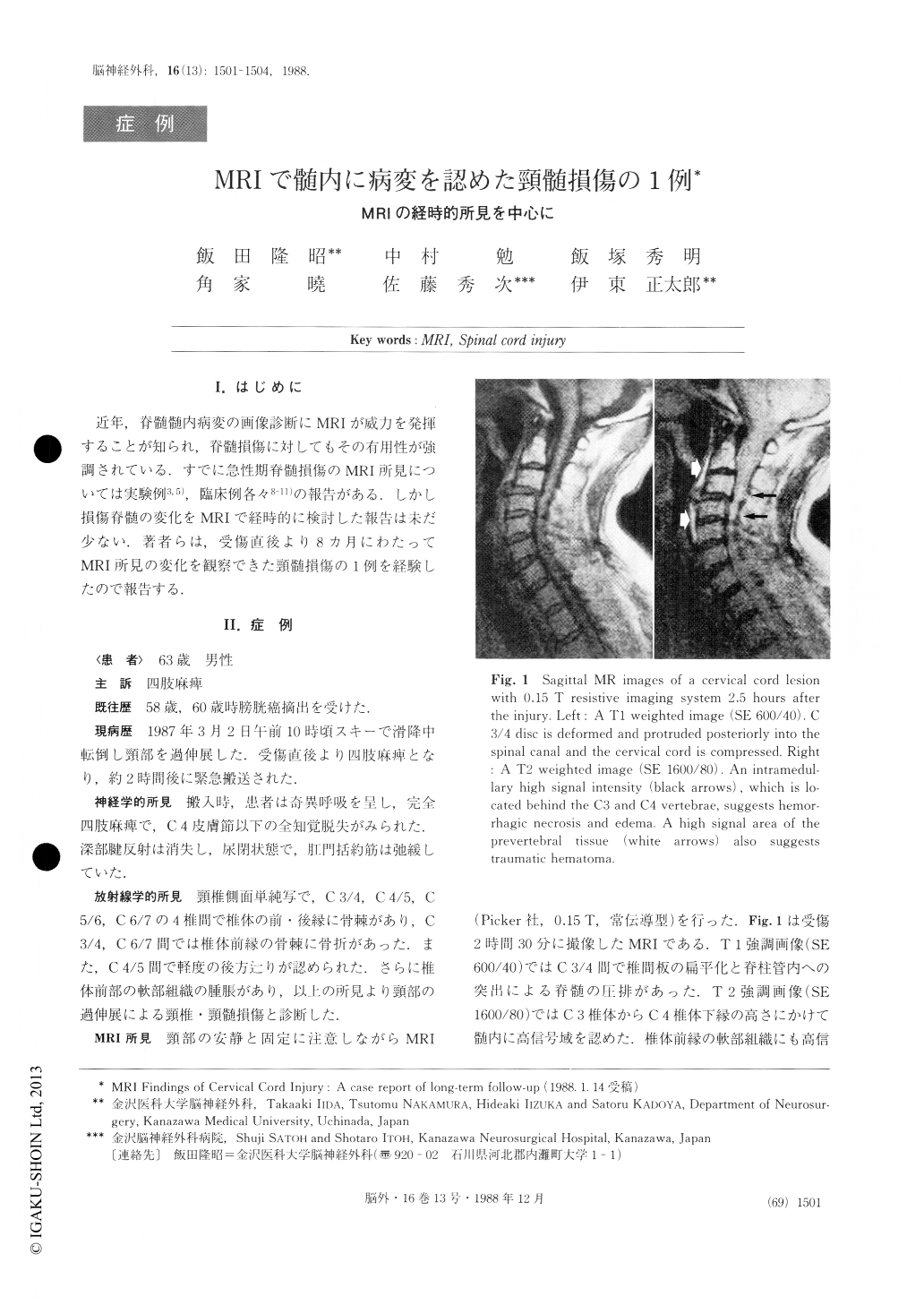
Right after the fall (2.5 hours later) the injured cord showed high intensity in T2 weighted images. This high intensity area stayed the same up to the chronic stage (6 months later). Ti weighted SE images showed no parenchymal change in the acute stage, but 3 months later a low intensity area appeared in the dam-aged cord.

Copyright © 1988, Igaku-Shoin Ltd. All rights reserved.


