Japanese
English
- 有料閲覧
- Abstract 文献概要
- 1ページ目 Look Inside
- 参考文献 Reference
Ⅰ.はじめに
近年,透析療法の進歩による慢性腎不全患者の増加により7),血液透析(hemodialysis:HD)療法中に脳神経疾患を生じ手術の必要性に迫られる機会が増加しているが,周術期の多岐にわたる全身合併症の存在により,未だ術中・術後管理に困難を伴うことが多い1,10).今回われわれは,HD患者に発生した髄膜腫の1症例を経験し,良好な経過を得ることができたので報告する.
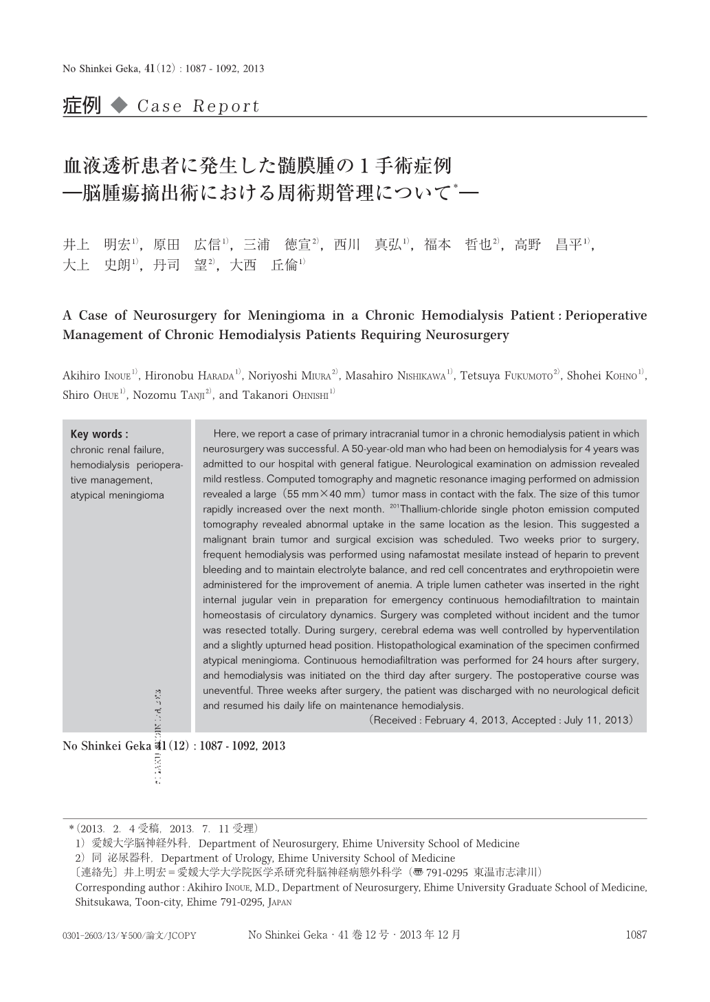
Here, we report a case of primary intracranial tumor in a chronic hemodialysis patient in which neurosurgery was successful. A 50-year-old man who had been on hemodialysis for 4 years was admitted to our hospital with general fatigue. Neurological examination on admission revealed mild restless. Computed tomography and magnetic resonance imaging performed on admission revealed a large(55mm×40mm)tumor mass in contact with the falx. The size of this tumor rapidly increased over the next month. 201Thallium-chloride single photon emission computed tomography revealed abnormal uptake in the same location as the lesion. This suggested a malignant brain tumor and surgical excision was scheduled. Two weeks prior to surgery, frequent hemodialysis was performed using nafamostat mesilate instead of heparin to prevent bleeding and to maintain electrolyte balance, and red cell concentrates and erythropoietin were administered for the improvement of anemia. A triple lumen catheter was inserted in the right internal jugular vein in preparation for emergency continuous hemodiafiltration to maintain homeostasis of circulatory dynamics. Surgery was completed without incident and the tumor was resected totally. During surgery, cerebral edema was well controlled by hyperventilation and a slightly upturned head position. Histopathological examination of the specimen confirmed atypical meningioma. Continuous hemodiafiltration was performed for 24hours after surgery, and hemodialysis was initiated on the third day after surgery. The postoperative course was uneventful. Three weeks after surgery, the patient was discharged with no neurological deficit and resumed his daily life on maintenance hemodialysis.

Copyright © 2013, Igaku-Shoin Ltd. All rights reserved.


