Japanese
English
- 有料閲覧
- Abstract 文献概要
- 1ページ目 Look Inside
- 参考文献 Reference
Ⅰ.はじめに
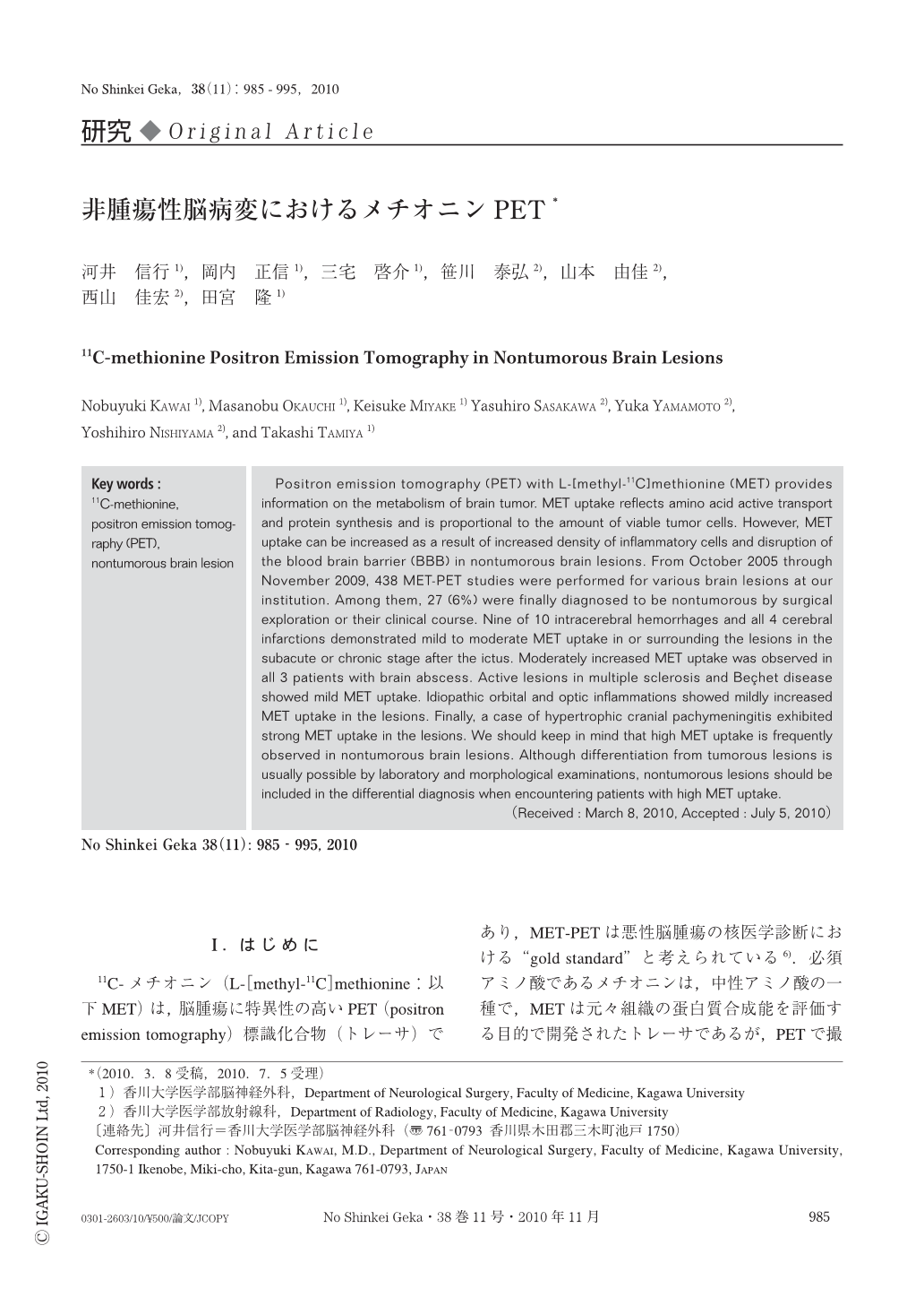
11C-メチオニン(L-[methyl-11C]methionine:以下MET)は,脳腫瘍に特異性の高いPET(positron emission tomography)標識化合物(トレーサ)であり,MET-PETは悪性脳腫瘍の核医学診断における“gold standard”と考えられている6).必須アミノ酸であるメチオニンは,中性アミノ酸の一種で,METは元々組織の蛋白質合成能を評価する目的で開発されたトレーサであるが,PETで撮影する時間内(投与後20~30分間)では組織へのアミノ酸輸送を主としてみていると考えられている.
METの組織への集積は,viableな腫瘍細胞に特異性が高く,非腫瘍性の脳病変にはMET集積が認められないことが報告されている2,12).METは局所での炎症反応の影響を18F-FDG(2-deoxy-2-[18F]fluoro-D-glucose:以下FDG)より受けにくいが7),マクロファージなどの炎症細胞や反応性に増殖したグリア細胞にもMET集積が認められる5).また投与されたMETは,主に中性アミノ酸輸送機構(L-type amino acid transporter 1)により血液脳関門(blood-brain barrier:以下BBB)を通して脳に能動的に取り込まれるが,一部は障害されたBBBを介した受動的な拡散もMET集積に関与している.そのため,非腫瘍性の脳病変においても局所の炎症反応やBBB破綻によりMET集積が認められ,ときに腫瘍性病変との鑑別が困難なことがある.
サイクロトロンを併設したPET施設の増加で,MET-PETが全国的に普及しはじめているが,METは腫瘍性脳病変以外でも陽性所見を示すことがあり,自験例を提示しながら概説し,注意を促したい.
Positron emission tomography (PET) with L-[methyl-11C]methionine (MET) provides information on the metabolism of brain tumor. MET uptake reflects amino acid active transport and protein synthesis and is proportional to the amount of viable tumor cells. However, MET uptake can be increased as a result of increased density of inflammatory cells and disruption of the blood brain barrier (BBB) in nontumorous brain lesions. From October 2005 through November 2009, 438 MET-PET studies were performed for various brain lesions at our institution. Among them, 27 (6%) were finally diagnosed to be nontumorous by surgical exploration or their clinical course. Nine of 10 intracerebral hemorrhages and all 4 cerebral infarctions demonstrated mild to moderate MET uptake in or surrounding the lesions in the subacute or chronic stage after the ictus. Moderately increased MET uptake was observed in all 3 patients with brain abscess. Active lesions in multiple sclerosis and Beçhet disease showed mild MET uptake. Idiopathic orbital and optic inflammations showed mildly increased MET uptake in the lesions. Finally, a case of hypertrophic cranial pachymeningitis exhibited strong MET uptake in the lesions. We should keep in mind that high MET uptake is frequently observed in nontumorous brain lesions. Although differentiation from tumorous lesions is usually possible by laboratory and morphological examinations, nontumorous lesions should be included in the differential diagnosis when encountering patients with high MET uptake.

Copyright © 2010, Igaku-Shoin Ltd. All rights reserved.


