Japanese
English
- 有料閲覧
- Abstract 文献概要
- 1ページ目 Look Inside
- 参考文献 Reference
Ⅰ.はじめに
M1 segmentが短い症例でのM1-2分岐部,あるいはearly bifurcation of the lateral orbitofrontal arteryの分岐部の動脈瘤は脳血管造影上,正面像で上方へprojectionし,解剖学的にはlimen insulaeの直下に存在する2,5).中大脳動脈瘤のうちM1の上方向きのものは,レンズ核線条体動脈の障害の可能性が他の部位,方向の中大脳動脈瘤に比べ高い6).しかし実際にはクリップで穿通枝を閉塞させるような初歩的なミスは少なく,M1の過度な牽引によりレンズ核線条体動脈領域の梗塞を生じたと思われる例,破裂動脈瘤での動脈瘤頚部の損傷,中枢確保前に瘤の操作を始めクリップをapplyしたが破裂部の閉鎖に難渋した例,などこの部位の動脈瘤の注意点はクリッピング操作そのものより,sylvian fissureの剝離範囲を含めた動脈瘤のexposureにあるのではないかと考えられる.
Yaşargilの著書,『Microneurosurgery』の中大脳動脈瘤の項目の中で,medial wall of proximal middle cerebral arteryに発生した14例について記述がある16).また,文献的にはM1部動脈瘤としてanterior temporal artery分岐部瘤を含めるものもあるが,手術戦略や難易度は異なり別個のものとして考えたほうがよいのではないかと思われた3,11,17).今回,leteral orbitofrontal arteryが早期に分岐しfalse bifurcationの形態をとるもので,この動脈分岐部から発生し結果としてmedial wallに発生したものを1つの範疇として検討した.この部位の動脈瘤は解剖学的にlimen recessに存在するが10,13),同部位の動脈瘤の自験例をretrospectiveに検討することにより,必要なmicrosurgical exposure,動脈瘤の処置に関して考察を行った.
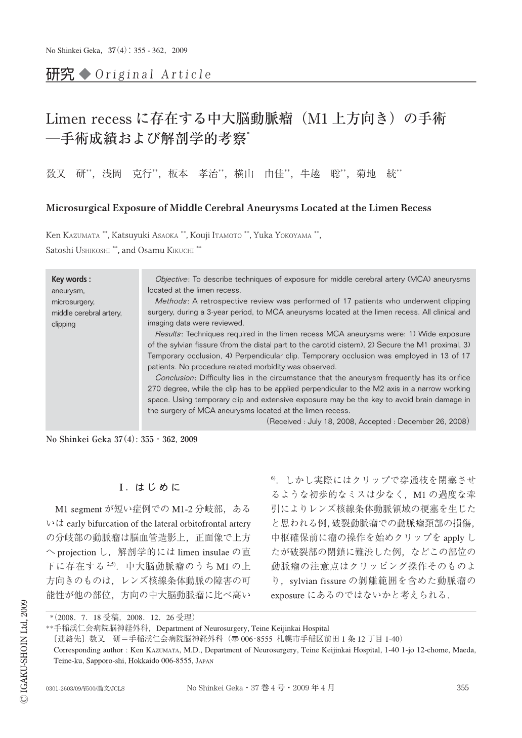
Objective: To describe techniques of exposure for middle cerebral artery (MCA) aneurysms located at the limen recess.
Methods: A retrospective review was performed of 17 patients who underwent clipping surgery, during a 3-year period, to MCA aneurysms located at the limen recess. All clinical and imaging data were reviewed.
Results: Techniques required in the limen recess MCA aneurysms were: 1) Wide exposure of the sylvian fissure (from the distal part to the carotid cistern), 2) Secure the M1 proximal, 3) Temporary occlusion, 4) Perpendicular clip. Temporary occlusion was employed in 13 of 17 patients. No procedure related morbidity was observed.
Conclusion: Difficulty lies in the circumstance that the aneurysm frequently has its orifice 270 degree, while the clip has to be applied perpendicular to the M2 axis in a narrow working space. Using temporary clip and extensive exposure may be the key to avoid brain damage in the surgery of MCA aneurysms located at the limen recess.

Copyright © 2009, Igaku-Shoin Ltd. All rights reserved.


