Japanese
English
- 有料閲覧
- Abstract 文献概要
- 1ページ目 Look Inside
- 参考文献 Reference
Ⅰ.緒言
頭蓋縫合早期癒合症は,1万人に1~5人に発生する44,45).頭蓋縫合早期癒合症のうち頭蓋冠および顔面骨に広範な形態異常を合併するものはsyndromic craniosynostosisに分類され7),重症例では頭蓋顔面奇形に対する乳児期からの多段階的手術や水頭症の管理,気管形成不全に対する呼吸管理などの集学的治療が必要である.今回,当院で経験した乳児期の頭蓋形成術,水頭症の管理,気管切開と人工呼吸器管理を要した最重症のsyndromic craniosynostosis 6例の治療と問題点につき報告する.
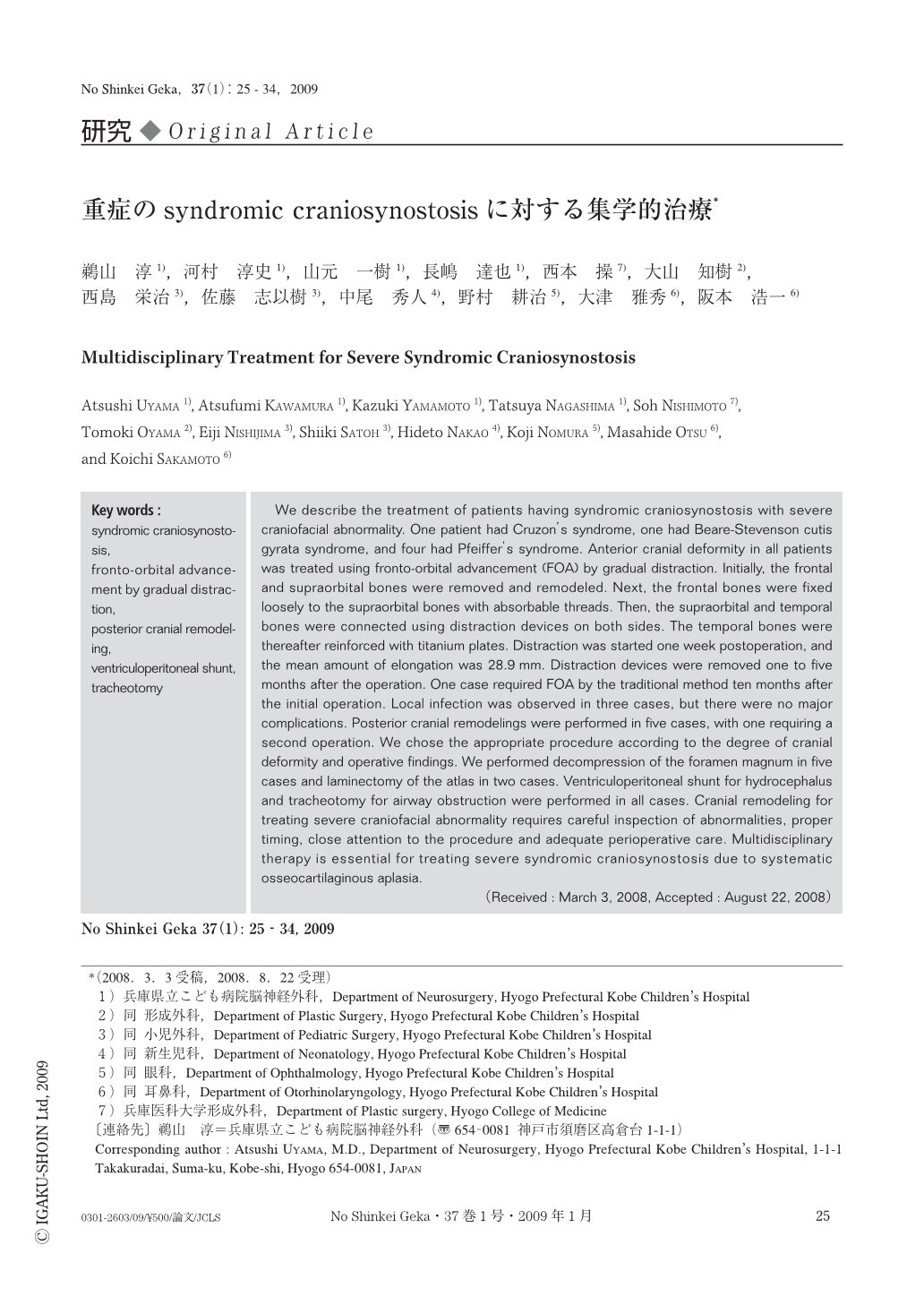
We describe the treatment of patients having syndromic craniosynostosis with severe craniofacial abnormality. One patient had Cruzon's syndrome, one had Beare-Stevenson cutis gyrata syndrome, and four had Pfeiffer's syndrome. Anterior cranial deformity in all patients was treated using fronto-orbital advancement (FOA) by gradual distraction. Initially, the frontal and supraorbital bones were removed and remodeled. Next, the frontal bones were fixed loosely to the supraorbital bones with absorbable threads. Then, the supraorbital and temporal bones were connected using distraction devices on both sides. The temporal bones were thereafter reinforced with titanium plates. Distraction was started one week postoperation, and the mean amount of elongation was 28.9mm. Distraction devices were removed one to five months after the operation. One case required FOA by the traditional method ten months after the initial operation. Local infection was observed in three cases, but there were no major complications. Posterior cranial remodelings were performed in five cases, with one requiring a second operation. We chose the appropriate procedure according to the degree of cranial deformity and operative findings. We performed decompression of the foramen magnum in five cases and laminectomy of the atlas in two cases. Ventriculoperitoneal shunt for hydrocephalus and tracheotomy for airway obstruction were performed in all cases. Cranial remodeling for treating severe craniofacial abnormality requires careful inspection of abnormalities, proper timing, close attention to the procedure and adequate perioperative care. Multidisciplinary therapy is essential for treating severe syndromic craniosynostosis due to systematic osseocartilaginous aplasia.

Copyright © 2009, Igaku-Shoin Ltd. All rights reserved.


