Japanese
English
- 有料閲覧
- Abstract 文献概要
- 1ページ目 Look Inside
Ⅰ.はじめに
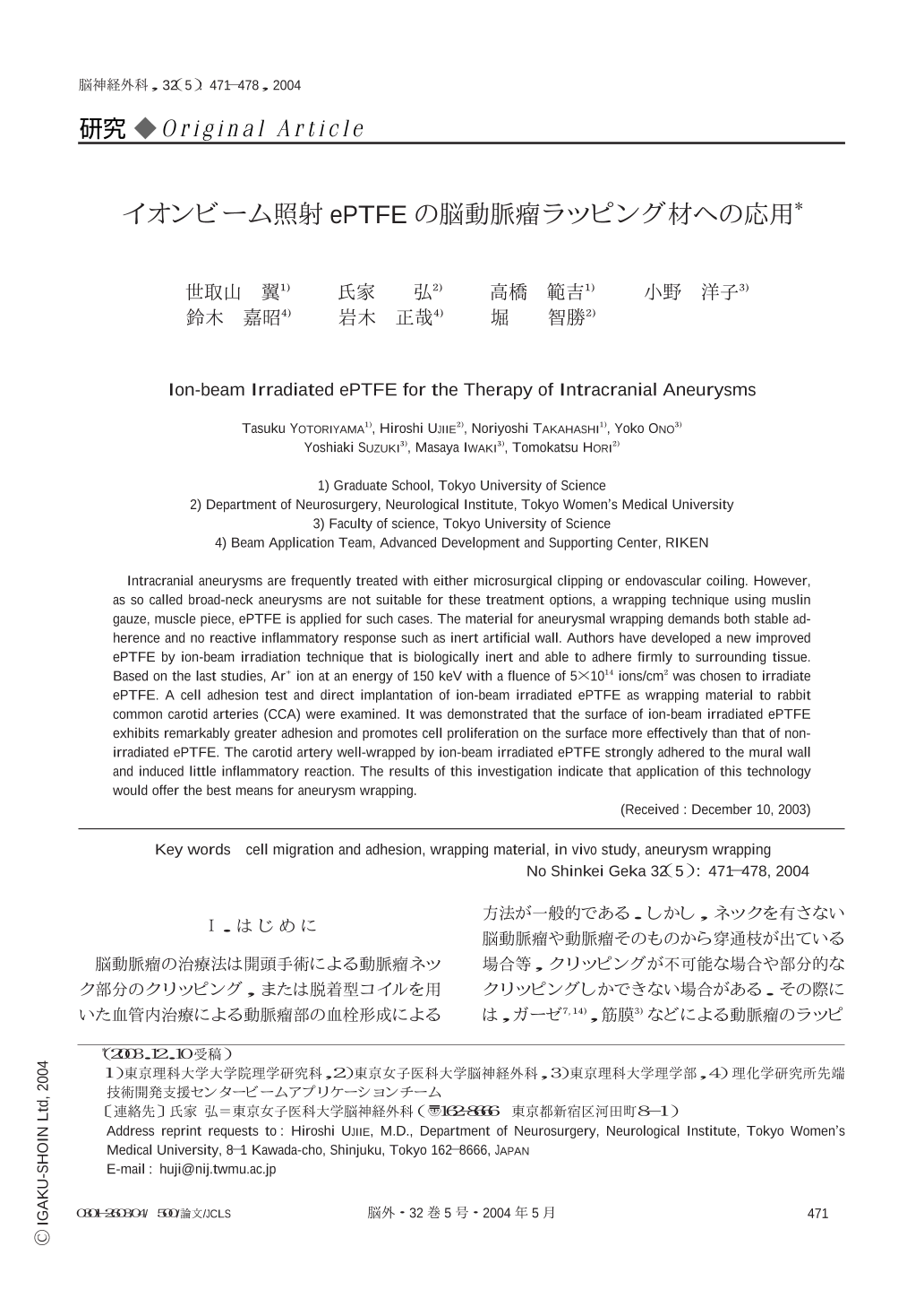
脳動脈瘤の治療法は開頭手術による動脈瘤ネック部分のクリッピング,または脱着型コイルを用いた血管内治療による動脈瘤部の血栓形成による方法が一般的である.しかし,ネックを有さない脳動脈瘤や動脈瘤そのものから穿通枝が出ている場合等,クリッピングが不可能な場合や部分的なクリッピングしかできない場合がある.その際には,ガーゼ7,14),筋膜3)などによる動脈瘤のラッピング後,フィブリン糊と呼ばれる生体組織接着剤による固定が行われる.しかし,筋膜は血管壁に対する密着性は優れているが吸収されてしまい永続的なラッピング材には適さず,また,綿ガーゼは操作性には優れているが強い異物反応,そして炎症性反応を示すため,ラッピング後に親動脈の狭窄および炎症性肉芽腫形成を起こす可能性が高い1,9).動脈瘤の理想的なラッピング材料の条件は,材料が生体内で異物反応,炎症反応,細胞毒性を起こさず,吸収されることなく永続的に動脈瘤壁に癒着し,動脈瘤の成長と破裂を防ぐことである.延伸ポリテトラフルオロエチレン(expanded polytetrafluoroethylene ; ePTFE)は,側鎖にフッ素を有する高分子素材であるため,生体内では組織との反応性が極めて低く,また構造的に極めて安定である.そのため,人工血管10,21),人工心膜2,6),人工腹膜12),人工腱索8),歯周組織再生誘導法における遮蔽膜4)などの生体材料として広く用いられている.
しかし,ePTFEは血管壁への親和性およびフィブリン糊の接着性が乏しく,現在までラッピング素材として使用されたことはない.
筆者らは,高分子ポリマー材料にイオンビームを照射することによって,その表層構造を改質し細胞接着性を付与することが可能であることを報告してきた15,18,19).ePTFEに関しても,Ne+を加速エネルギー150 keVで1×10¹⁵ ions/cm2照射したePTFEが,細胞接着性を付与されることを見出した.さらにウサギ頭蓋骨および背部筋膜上への留置実験によって,骨および筋膜組織との接着性を有することを明らかにした16,17,20).また,加速エネルギー150 keVでAr+,Kr+イオンビーム照射したePTFE表面も同様に細胞接着性を有し,フィブリン糊との強力な接着を有することも明らかになった22).
本研究では,in vitroで細胞培養実験を行い,加えてイオンビーム照射後のePTFE表面を走査型電子顕微鏡で評価し,イオンビーム照射したePTFEシートが動脈瘤ラッピング材料として有効な素材になり得るか検討した.
今までの基礎実験の結果15-20)から,加速エネルギー150 keVでAr+イオンを5×10¹⁴ ions/cm2照射したePTFEと未照射のePTFEを用いて,ウサギの頸動脈を用いたラッピング実験を行って検討した.
Intracranial aneurysms are frequently treated with either microsurgical clipping or endovascular coiling. However,as so called broad-neck aneurysms are not suitable for these treatment options,a wrapping technique using muslin gauze,muscle piece,ePTFE is applied for such cases. The material for aneurysmal wrapping demands both stable adherence and no reactive inflammatory response such as inert artificial wall. Authors have developed a new improved ePTFE by ion-beam irradiation technique that is biologically inert and able to adhere firmly to surrounding tissue. Based on the last studies,Ar+ ion at an energy of 150 keV with a fluence of 5×10¹⁴ ions/cm2 was chosen to irradiate ePTFE. A cell adhesion test and direct implantation of ion-beam irradiated ePTFE as wrapping material to rabbit common carotid arteries (CCA) were examined. It was demonstrated that the surface of ion-beam irradiated ePTFE exhibits remarkably greater adhesion and promotes cell proliferation on the surface more effectively than that of non-irradiated ePTFE. The carotid artery well-wrapped by ion-beam irradiated ePTFE strongly adhered to the mural wall and induced little inflammatory reaction. The results of this investigation indicate that application of this technology would offer the best means for aneurysm wrapping.

Copyright © 2004, Igaku-Shoin Ltd. All rights reserved.


