Japanese
English
- 有料閲覧
- Abstract 文献概要
- 1ページ目 Look Inside
I.はじめに
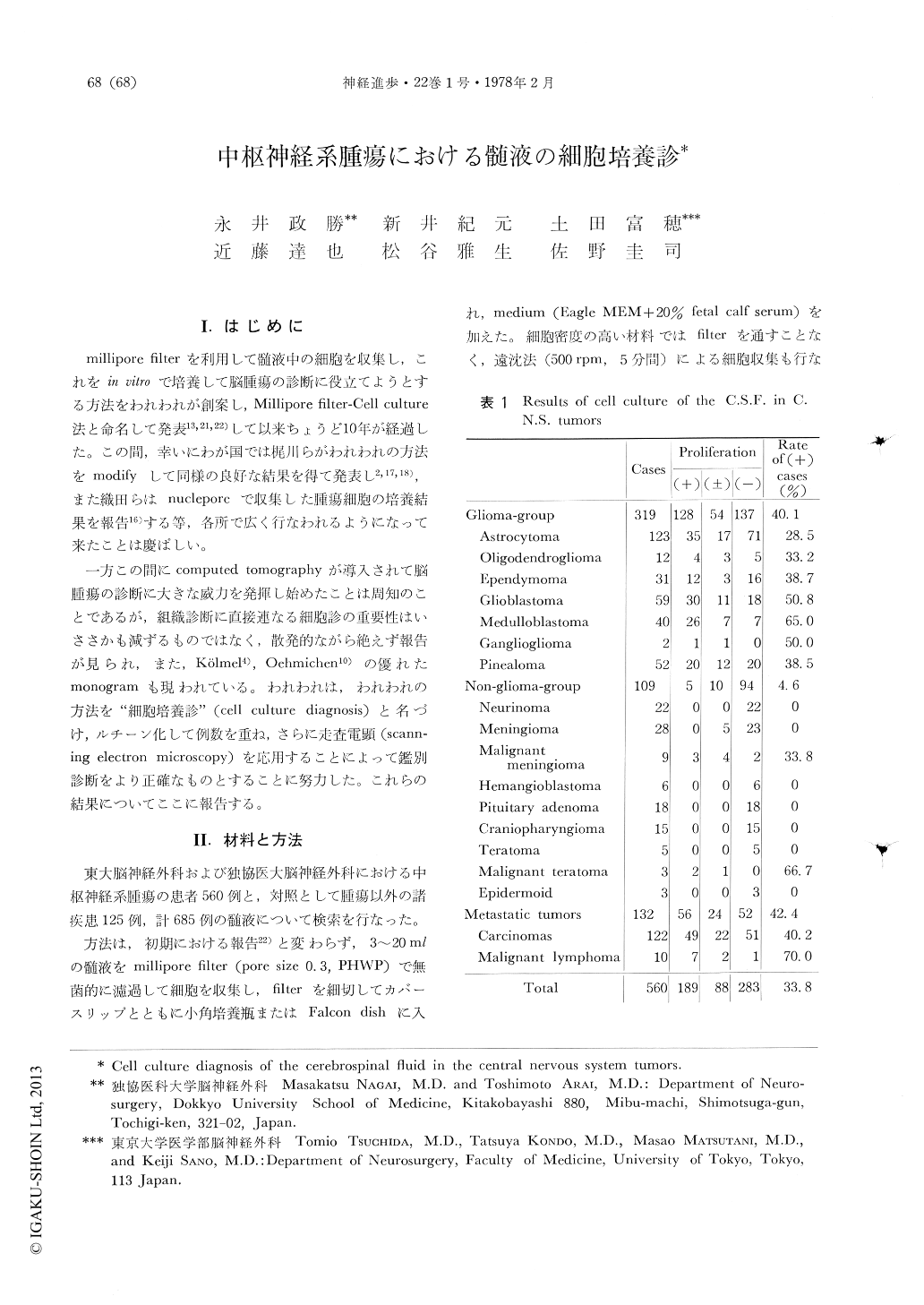
millipore filterを利用して髄液中の細胞を収集し,これをin vitroで培整して脳腫瘍の診断に役立てようとする方法をわれわれが創案し,Millipore filter-Cell culture法と命名して発表13,21,22)して以来ちょうど10年が経過した。この間,幸いにわが国では梶川らがわれわれの方法をmodifyして同様の良好な結果を得て発表し2,17,18),また織田らはnucleporeで収集した腫瘍細胞の培養結果を報告16)する等,各所で広く行なわれるようになって来たことは慶ばしい。
一方この間にcomputed tomographyが導入されて脳腫瘍の診断に大きな威力を発揮し始めたことは周知のことであるが,組織診断に直接連なる細胞診の重要性はいささかも減ずるものではなく,散発的ながら絶えず報告が見られ,また,Kölmel4),Ochmichen10)の優れたmonogramも現われている。われわれは,われわれの方法を"細胞培養診"(cell culture diagnosis)と名づけ,ルチーン化して例数を重ね,さらに走査電顕(scanning electron microscopy)を応用することによって鑑別診断をより正確なものとすることに努力した。これらの結果についてここに報告する。
The authors reported ten years ago, a new diagnostic method of the cerebrospinal fluid in central nervous system tumors. The cells were collected with millipore filter and cultured for several days. The diagnosis was made from the proliferating pattern of the cultured cells comparing with that of the trypsinization-monolayer culture of the tumor tissue itself. 560 cases of the central nervous system tumor were examined bythis method and 189 cases showed proliferations in vitro (positive cases) : Glioma group-126/319, 40.1%, non-glioma group -5/109, 4.6%, metastatic tumors -56/132, 42.4%.

Copyright © 1978, Igaku-Shoin Ltd. All rights reserved.


