Japanese
English
- 有料閲覧
- Abstract 文献概要
- 1ページ目 Look Inside
I.はじめに
従来,私どもは一連の糖脂質の化学的研究とともに免疫化学的研究を行なつてきているが,今回,これら糖脂質の組織分布を解明せんとして,螢光抗体法による組織化学的研究を試みてみた。もちろん,糖脂質の免疫化学的研究自体まだ問題を残しているが,最近ではRapport一派1)2)やKuwert,Niedieck,Pette3)4)5),その他6)7)によつて中枢神経系の組織特異抗原性がセレブロシドに由来するものであるという報告が注目されている。
私どもはこれらとはまつたく独立に糖脂質の免疫化学的研究を進めてきた。脂質は水不溶のため免疫学的研究の対象として不利であり,従来,脂質特異抗体を沈降反応によつて証明することはできなかつた。脂質のこれらの欠陥を補うため,一種の人工リポ蛋白を合成して,化学構造の明確な脂質部分に特異的な抗体が得られないか,しかも定量沈降反応によつて認められる抗体が得られないかと追究した。
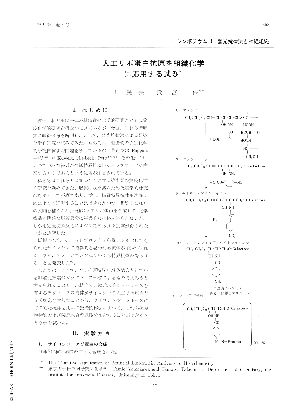
We attempted to prepare artificial antigens capable of producing by themselves the lipid hapten-specific antibodies. At first, a synthetic psychosine-protein was prepared as follows; the psychosine (sphingosyl-galactoside) was obtained by the alkaline hydrolysis of cerebroside. The synthetic N-p-, minobenzoyldihydropsychosine was conjugatedwith bovine serum albumin or egg albumin byth diszo-coupling reaction. Secondly, according to Babers & Goebel, we prepared an azo proteincontaining a lactose as a determinant group.
These artificial antigens as alum precipitates were injected subcutaneously or intravenously to rabbits to get immune sera. After the assay of psychosine- or lactose-specific antibodies by the qualitative and quantitative precipitin tests and the Ouchterlony method, these immune sera wereapplied to the fluorescent antibody technique to understand the distribution of cerebroside or its related glycolipids in various tissues. In thepresent paper, brain and kidney tissues from the mouse were frozen in hexane chilled to -70℃. Sections were cut at 4p in a cryostat at -20℃. The staining of the freshly frozen tissue sectionwith fluorescent anti-psychosine or anti-lactose solution was carried out without prior fixation or with treatment of organic solvents. When sections of mouse brain were stained with fluorescent antibody to psychosine or lactose, the fluorescence appeared consistently in the lumen of the cerebral blood vessel and in the plexus, but not in the myelinsheath of white matter contrary to our expectation. In Kctions from kidney stained with fluorescentantibodies, the tubules and the renal blood vesselshowed the brilliant fluorescence, but the glomerulus did not show.
The localization of the glycolipids in the mouse brain and kidney by means of fluorescent antibody technique was discussed briefly in view of these results.

Copyright © 1965, Igaku-Shoin Ltd. All rights reserved.


