Japanese
English
- 有料閲覧
- Abstract 文献概要
- 1ページ目 Look Inside
- 参考文献 Reference
はじめに
近年,若年者の脳卒中に関連して脳動脈解離が注目されている。欧米では古くから脳梗塞の原因として頸部頸動脈解離の存在が知られていたが,1977年Yonasらにより頭蓋内の動脈解離が報告され1),本邦では1995年の脳神経外科施設を対象とした調査により,若年者くも膜下出血の原因として広く認識されるようになった2,3)。脳動脈解離は脳梗塞やくも膜下出血という異なった臨床症状を呈し,また画像所見が経過中に変化しうるなど病態が複雑で不明な点が多い4,5)。それゆえ治療法に関してのエビデンスがなく,明確な指針が示されていないのが現状である。
脳動脈解離は,頭蓋内動脈を指す(cerebral arterial dissection)場合と,頸部頸動脈・椎骨動脈より末梢の頭蓋外・頭蓋内動脈を広く指す(cervicocephalic arterial dissection)場合とがあるが,一般には後者の概念が脳を灌流する動脈の解離として受け入れられている5)。また外傷性(traumatic)と特発性(spontaneous)に分類されるが,ここでは特発性脳動脈解離について明らかとなっている事項を解説する。
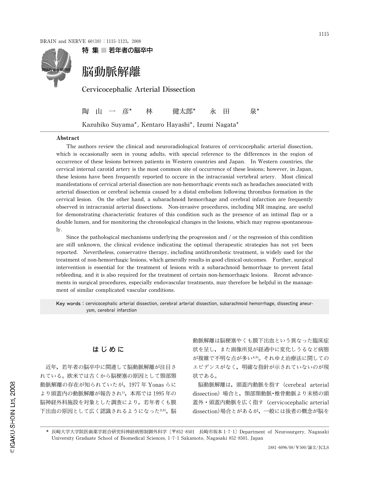
Abstract
The authors review the clinical and neuroradiological features of cervicocephalic arterial dissection,which is occasionally seen in young adults,with special reference to the differences in the region of occurrence of these lesions between patients in Western countries and Japan. In Western countries,the cervical internal carotid artery is the most common site of occurrence of these lesions; however,in Japan,these lesions have been frequently reported to occure in the intracranial vertebral artery. Most clinical manifestations of cervical arterial dissection are non-hemorrhagic events such as headaches associated with arterial dissection or cerebral ischemia caused by a distal embolism following thrombus formation in the cervical lesion. On the other hand,a subarachnoid hemorrhage and cerebral infarction are frequently observed in intracranial arterial dissections. Non-invasive procedures,including MR imaging,are useful for demonstrating characteristic features of this condition such as the presence of an intimal flap or a double lumen,and for monitoring the chronological changes in the lesions,which may regress spontaneously.
Since the pathological mechanisms underlying the progression and / or the regression of this condition are still unknown,the clinical evidence indicating the optimal therapeutic strategies has not yet been reported. Nevertheless,conservative therapy,including antithrombotic treatment,is widely used for the treatment of non-hemorrhagic lesions,which generally results in good clinical outcomes. Further,surgical intervention is essential for the treatment of lesions with a subarachnoid hemorrhage to prevent fatal rebleeding,and it is also required for the treatment of certain non-hemorrhagic lesions. Recent advancements in surgical procedures,especially endovascular treatments,may therefore be helpful in the management of similar complicated vascular conditions.

Copyright © 2008, Igaku-Shoin Ltd. All rights reserved.


