Japanese
English
- 有料閲覧
- Abstract 文献概要
- 1ページ目 Look Inside
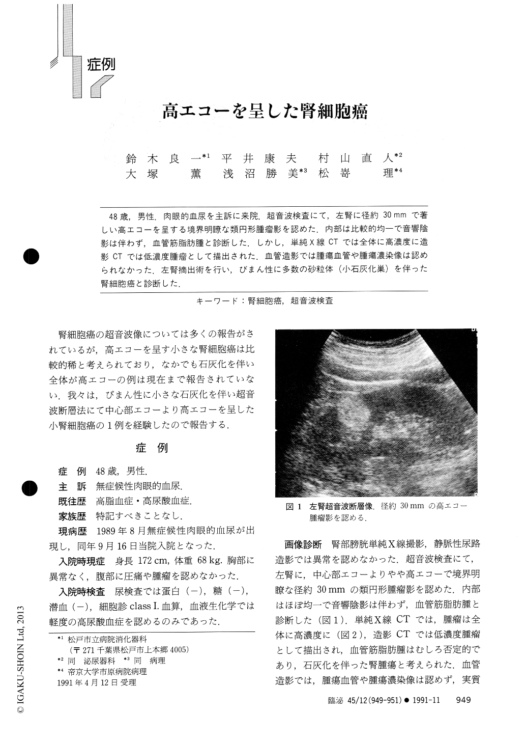
48歳,男性.肉眼的血尿を主訴に来院.超音波検査にて,左腎に径約30mmで著しい高エコーを呈する境界明瞭な類円形腫瘤影を認めた.内部は比較的均一で音響陰影は伴わず,血管筋脂肪腫と診断した.しかし,単純X線CTでは全体に高濃度に造影CTでは低濃度腫瘤として描出された.血管造影では腫瘍血管や腫瘍濃染像は認められなかった.左腎摘出術を行い,びまん性に多数の砂粒体(小石灰化巣)を伴った腎細胞癌と診断した.
A case with hyperechoic renal cell carcinoma, indicating that the presence of a hyperechoic renal mass is not pathognomonic for angiomyolipoma, was described. A 48-year-old man was hospitalized with a complaint of grosshematuria. Ultrasonotomography demonstrated a homogeneously hyperechoic mass show-ing 30 mm in diameter in the left kidney. The level of internal echo was as echogenic as that of the central echo complex. The CT density of the mass, however, was not low but high. Gross appearance of the surgically removed tumor was 30 mm in the greatest diameter with the cut surface appearing tan-brown without bleeding and necrosis.

Copyright © 1991, Igaku-Shoin Ltd. All rights reserved.


