Japanese
English
- 有料閲覧
- Abstract 文献概要
- 1ページ目 Look Inside
- 参考文献 Reference
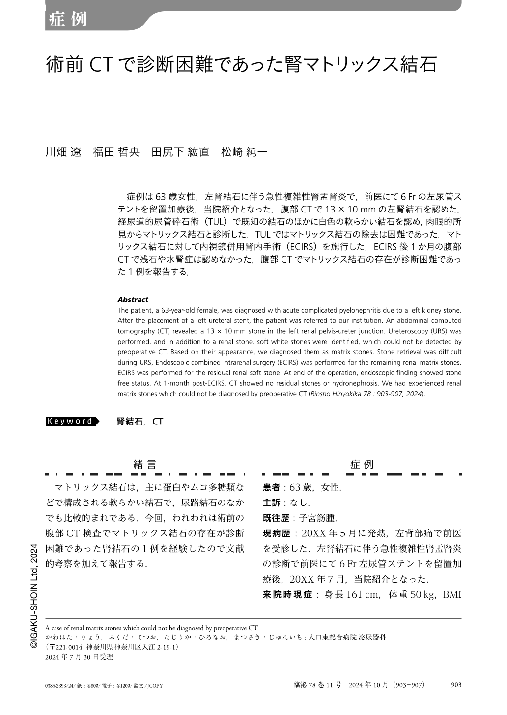
症例は63歳女性.左腎結石に伴う急性複雑性腎盂腎炎で,前医にて6Frの左尿管ステントを留置加療後,当院紹介となった.腹部CTで13×10mmの左腎結石を認めた.経尿道的尿管砕石術(TUL)で既知の結石のほかに白色の軟らかい結石を認め,肉眼的所見からマトリックス結石と診断した.TULではマトリックス結石の除去は困難であった.マトリックス結石に対して内視鏡併用腎内手術(ECIRS)を施行した.ECIRS後1か月の腹部CTで残石や水腎症は認めなかった.腹部CTでマトリックス結石の存在が診断困難であった1例を報告する.
Abstract
The patient, a 63-year-old female, was diagnosed with acute complicated pyelonephritis due to a left kidney stone. After the placement of a left ureteral stent, the patient was referred to our institution. An abdominal computed tomography (CT) revealed a 13 × 10mm stone in the left renal pelvis-ureter junction. Ureteroscopy (URS) was performed, and in addition to a renal stone, soft white stones were identified, which could not be detected by preoperative CT. Based on their appearance, we diagnosed them as matrix stones. Stone retrieval was difficult during URS, Endoscopic combined intrarenal surgery (ECIRS) was performed for the remaining renal matrix stones. ECIRS was performed for the residual renal soft stone. At end of the operation, endoscopic finding showed stone free status. At 1-month post-ECIRS, CT showed no residual stones or hydronephrosis. We had experienced renal matrix stones which could not be diagnosed by preoperative CT (Rinsho Hinyokika 78 : 903-907, 2024).

Copyright © 2024, Igaku-Shoin Ltd. All rights reserved.


