Japanese
English
- 有料閲覧
- Abstract 文献概要
- 1ページ目 Look Inside
いわゆる若年性黒色腫juvenile melanoma(以下JM)は,Spitz1)(1948)以前には小児のmalignant melanoma (以下MM)と考えられていた。本症の名称はSpitzの命名せるJMが広く用いられてはいるが,論議のあるところで,したがつて,数多くの同義語がある。これを列挙すると,Naevus prominens et pigmentosus(Du Bois2)1934),spindle cell and epithelioid cell nevus(Helwig3)1954),pseudomélanome (Grupper et Tubiana4) 1955),benign juvenile melanoma(Kopf and Andrade5) 1954),Mc-Whorter and Woolner6)1954, Steigleder u. Wellmer7) 1956, Lever8)1967, Montgomery9) 1967,石川10)1969),tumeur de Spitz(Du- perrat et Mascaro11) 1961),Spindelzellen-, Epitheloidzellen-oder gemischter Spindel Epitheloid-zellennävus(Gartmann12) 1962), Naevus Spitz(Cottini13) 1963),nevus with large cells(Duverne et Prunieras14) 1965), Blasenzellnaevus(Schauer u.Vogel15) 1967), juvenile pseudomelanoma(小嶋ら16) 1968), mélanome de Spitz(Degos17) 1964, Fischer18) 1968, Hadida et al.19) 1969),Sourreil et al.20) 1969),melanocytic nevus, Spitz nevus(Mc-Govern and Brown21)1969)など枚挙にいとまないが,アメリカではbenign juvenile mela-noma,ドイツではsog juveniles Melanom,フランスではmélanome de Spitzないしtu-meur de Spitzが多く用いられているようである。名称に関する問題点は,成人例がかなりあるのに"juvenile"が,また悪性ではないのに"melanoma"が用いられていることにある。Degos17)は,"mélanome"という語は黒い腫瘍という意味でもなければ,悪性という意味でもないからmélanome de Spitzがよいとしているが,"melanoma"はAmerican Medical Associa-tion's Standard Nomenclatureによれば,malignant lesionに対する公式の用語であり,すなわちMMを意味する(Kopt and Andra-de5))。したがつて,histogenesisがより明確になるまでは,暫定的にSpitz nevusあるいはtu-meur de Spitzとしておいた方が妥当かもしれない(McGovern and Brown21))。
近年,本邦においても本症の報告例は増加してきており,現在までに57例を数えるに至つた。著者らは,最近,本症の3例を経験したので若干の文献的考察とともに報告する。
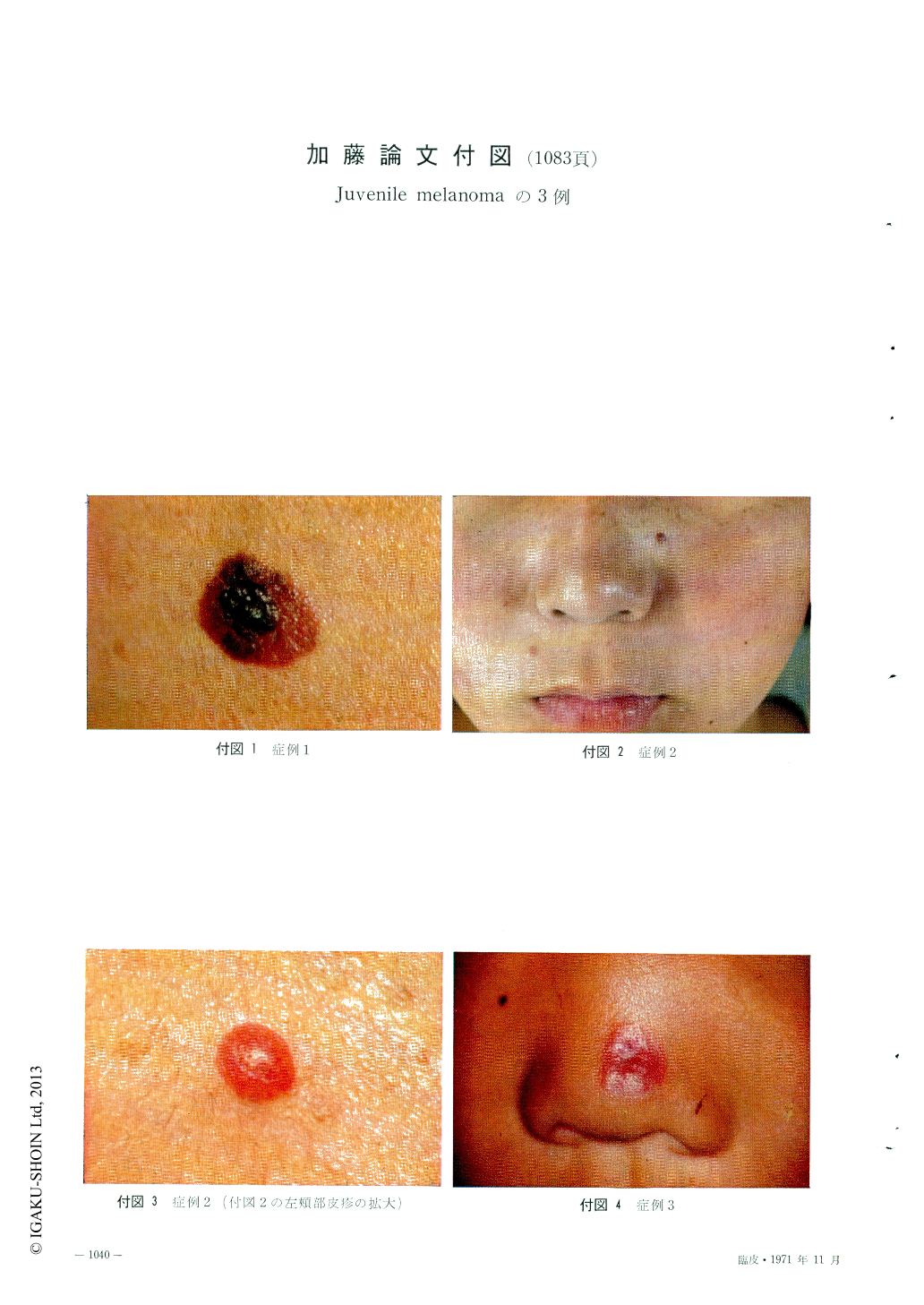
Three cases of Juvenile melanoma on the face in 16, and 13 year-old girls and 5 year-old boy showed clinically typical picture, but histologically the second and third case showed atypical picture partly looks like the mesenchymal tumor. Statistical studies on 57 cases reported in Japan were performed. There was no sex difference in the frequency. Average age of the first visit was 12.2, ranging from 24 hours to 50 years of age. Cases under 15-year-old were 72.2%. Predilection site was the cheek, where almost all cases of the face were found, and then nose. 85.2% of all cases were found on the face.
Almost all cases had a solitary lesion except 3 cases. In spite of the detailed diagnostic histo-logical criteria, histological pictures seemed to have considerable varieties.

Copyright © 1971, Igaku-Shoin Ltd. All rights reserved.


